

Lower diffusion in white matter of children with prenatal methamphetamine exposure
- PMID: 19369643
- PMCID: PMC2697962
- DOI: 10.1212/01.wnl.0000346516.49126.20
Lower diffusion in white matter of children with prenatal methamphetamine exposure
Abstract
Background: Methamphetamine use is a common problem among women of childbearing age, leading to an increasing number of children with prenatal methamphetamine exposure. Whether microstructural brain changes associated with prenatal methamphetamine exposure can be detected with diffusion tensor imaging (DTI) is unknown.
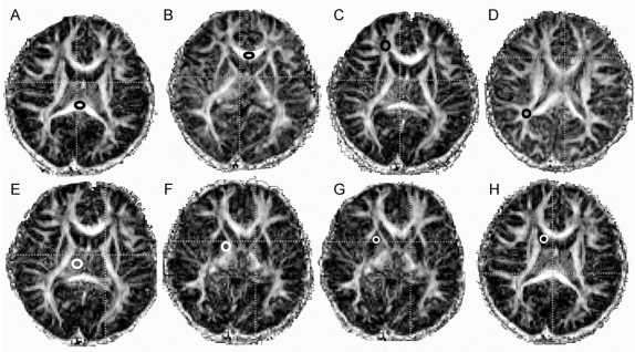
Method: Twelve-direction DTI was performed in 29 methamphetamine-exposed and 37 unexposed children ages 3-4 years on a 3-T MRI scanner. Fractional anisotropy (FA) and apparent diffusion coefficient (ADC) were determined in the corpus callosum (genu and splenium) and bilaterally in the frontal and parietal white matter (WM), basal ganglia (caudate, putamen, globus pallidus), and thalamus.
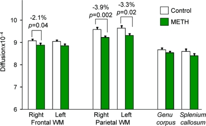
Results: Children with prenatal methamphetamine exposure had lower ADC in the frontal (right: -2.1%, p = 0.04; left: -2.0%, p = 0.09) and parietal WM (right: -3.9%, p = 0.002; left: -3.3%, p = 0.02) compared to unexposed children. The methamphetamine-exposed children also showed a trend for higher FA in the left frontal WM (+4.9%, p = 0.06) compared to the unexposed children.
Conclusion: Since less myelination and higher dendritic or spine density have been reported in animals exposed to methamphetamine, lower diffusion in our children may reflect more compact axons or greater dendritic or spine density associated with prenatal methamphetamine exposure. These findings suggest alterations in white matter maturation in these children exposed to methamphetamine in utero.
Figures
Comment in
-
Still no time for complacency: Developmental effects of prenatal methamphetamine exposure.Neurology. 2009 Jun 16;72(24):2062-3. doi: 10.1212/WNL.0b013e3181a92c98. Epub 2009 Apr 29. Neurology. 2009. PMID: 19403882 No abstract available.
References
-
- Chang L, Smith LM, LoPresti C, et al. Smaller subcortical volumes and cognitive deficits in children with prenatal methamphetamine exposure. Psychiatry Res 2004;132:95–106. - PubMed
-
- Smith LM, Chang L, Yonekura ML, Grob C, Osborn D, Ernst T. Brain proton magnetic resonance spectroscopy in children exposed to methamphetamine in utero. Neurology 2001;57:255–260. - PubMed
-
- Unis AS, Roberson MD, Robinette R, Ha J, Dorsa DM. Ontogeny of human brain dopamine receptors: I: differential expression of [3H]-SCH23390 and [3H]-YM09151-2 specific binding. Brain Res Dev Brain Res 1998;106:109–117. - PubMed
-
- Zhang L, Lidow MS. D1 dopamine receptor regulation of cell cycle in FGF- and EGF-supported primary cultures of embryonic cerebral cortical precursor cells. Int J Dev Neurosci 2002;20:593–606. - PubMed
-
- Herlenius E, Lagercrantz H. Development of neurotransmitter systems during critical periods. Exp Neurol 2004;190 suppl 1:S8–21. - PubMed
Publication types
MeSH terms
Substances
Grants and funding
LinkOut - more resources
Full Text Sources
Medical