. 2009 Apr;87(4):287-99.
doi: 10.1139/Y09-010.

Involvement of UTP in protection of cardiomyocytes from hypoxic stress

Affiliations

Involvement of UTP in protection of cardiomyocytes from hypoxic stress

Asher Shainberg et al. Can J Physiol Pharmacol. 2009 Apr.

Abstract

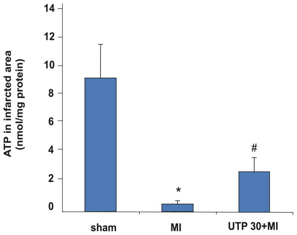
Massive amounts of nucleotides are released during ischemia in the cardiovascular system. Although the effect of the purine nucleotide ATP has been intensively studied in myocardial infarction, the cardioprotective role of the pyrimidine nucleotide UTP is still unclear, especially in the cardiovascular system. The purpose of our study was to elucidate the protective effects of UTP receptor activation and describe the downstream cascade for the cardioprotective effect. Cultured cardiomyocytes and left anterior descending (LAD)-ligated rat hearts were pretreated with UTP and exposed to hypoxia-ischemia. In vitro experiments revealed that UTP reduced cardiomyocyte death induced by hypoxia, an effect that was diminished by suramin. UTP caused several effects that could trigger a cardioprotective response: a transient increase of [Ca2+]i, an effect that was abolished by PPADS or RB2; phosphorylation of the kinases ERK and Akt, which was abolished by U0126 and LY294002, respectively; and reduced mitochondrial calcium elevation after hypoxia. In vivo experiments revealed that UTP maintained ATP levels, improved mitochondrial activity, and reduced infarct size. In conclusion, UTP administrated before ischemia reduced infarct size and improved myocardial function. Reduction of mitochondrial calcium overload can partially explain the protective effect of UTP after hypoxic-ischemic injury.


Figures

Fig. 1
Fig. 1
Concentration- and time-dependent effects of UTP on cultured cardiomyocytes subjected to hypoxia. (A) Six-day-old rat cardiomyocytes were treated for 15 min with various concentrations of UTP (3–50 μmol/L). The cells were then washed twice and subjected to hypoxia for 2 h in glucose-free PBS at 37 °C. The amount of lactate dehydrogenase (LDH) released to the medium was determined and compared with the total activity of control homogenate (100%). (B) Cardiomyocytes were treated for 15 min or 24 h with 50 μmol/L UTP before hypoxia. Other groups of cells were incubated for 1 h with 50 μmol/L UTP, washed and replaced in normal medium, and then subjected to hypoxia 24, 48, or 72 h subsequent to the treatment with UTP. Data are means of at least 3 replicates in 5 separate experiments ± SD. *, Significant at p < 0.05, **, p < 0.01 compared with hypoxia.
Fig. 2
Fig. 2
Effect of UTP on cardiomyocyte morphology under hypoxic conditions. Six-day-old rat cardiomyocytes cultured under normoxic conditions (A) were subjected to 2 h in an hypoxic environment (B) or pretreated with 50 μmol/L UTP and then subjected to hypoxia (C). Damaged cells were increased by hypoxia, whereas activation of UTP receptors before hypoxia reduced the percentage of cell death. Cells in the left column were stained with propidium iodide, which marks damaged cells red (medium grey in the print version, red in the Web version), and Hoechst 33342, which stains live-cell nuclei blue (lighter grey in the print version). Cells in the middle column were stained with hematoxylin and eosin, and those in the right column were stained with neutral red, which stains lysosomes. The results shown are representative of 6 experiments. Scale bars represent 10 μm.
Fig. 3
Fig. 3
Effect of P2 receptor agonists and antagonists on intracellular ATP levels after hypoxia in vitro. Six-day-old rat cardiomyocytes were treated for 15 min with the P2 antagonists PPADS (10 μmol/L), RB2 (50 μmol/L), or suramin (300 μmol/L) before application of UTP (50 μmol/L) for 15 min. Intracellular ATP levels were measured after 2 h of hypoxia. Data are means of at least 3 replicates in 6 separate experiments ± SD. *, Significant at p < 0.01 compared with hypoxia; #, p < 0.001 compared with normoxia.
Fig. 4
Fig. 4
Effect of UTP on [Ca2+]i accumulation during hypoxia. Effect of hypoxia alone (A) and 30 min (B) or 24 h (C) after UTP treatment for 15 min in 6-day-old rat cardiomyocytes in vitro. Calcium was monitored during hypoxia with fluorescent dye indo-1. The fluorescence ratio of 410/490 nm, which is proportional to changes in Ca2+ levels, is demonstrated. Each recording was made for 10 s at times 0, 20, 40, and 60 min after the initiation of hypoxia. Each experiment shown is representative of 5 separate experiments. (D) Basal levels of [Ca2+]i of the 3 groups and a normoxic control as a function of time. Values are means ± SD. *, Significant at p < 0.05 UTP-treated (30 min) compared with hypoxia.
Fig. 5
Fig. 5
Effect of pretreatment with UTP on mitochondrial Ca2+ overload in cardiomyocytes subjected to hypoxia. Six-day-old rat cardiomyocytes were treated with 50 μmol/L UTP 30 min or 24 h before 2 h of hypoxia. During hypoxia, cardiomyocytes were loaded with rhod-2 AM (warm after cold incubation), and Ca2+ level was determined. Values are means ± SD. *, Significant at p < 0.001 compared with control; #, p < 0.001 compared with hypoxia.
Fig. 6
Fig. 6
Effect of P2 antagonists on [Ca2+]i response to UTP. Indo-1-loaded 6-day-old rat cardiomyocytes were treated with 50 μmol/L UTP alone (A) or pretreated with the P2 antagonists PPADS (B), RB2 (C), or suramin (D) 15 min before UTP application. Other cells were pretreated before UTP with (E) IP3 receptor inhibitor 2APB (50 μmol/L) or (F) PLC inhibitor U73122 (2 μmol/L). The fluorescence ratio of 410/490 nm, which is proportional to changes in Ca2+ levels, is demonstrated. Each experiment shown is representative of 7 separate experiments. IP3, inositol 1,4,5-trisphosphate; 2APB, 2-aminoethoxydiphenyl borate; PLC, phospholipase C.
Fig. 7
Fig. 7
Time-dependent effects of UTP on ERK phosphorylation in 6-day-old rat cardiomyocytes. (A) Cells were treated with 50 μmol/L UTP for the indicated time. (B) Cells were treated with 50 μmol/L UTP for 5 min with or without 10 μmol/L U0126, a selective MEK1/2 inhibitor. Cell lysates were analyzed by Western blotting using a phosphospecific ERK antibody (Tyr204). The same samples were also analyzed on a separate blot using an antibody that recognizes desmin (total) to confirm equal loading on each lane. The combined ratio results (lower panel), obtained from densitometric analysis of blots, are representative of 3 identical independent experiments. Values are a percentage of the basal level of ERK phosphorylation (100%).
Fig. 8
Fig. 8
Effect of ERK and Akt inhibitors on UTP-induced cardioprotection. Six-day-old rat cardiomyocytes were treated for 15 min with 10 μmol/L U0126 (a selective inhibitor of MEK1/2) and 30 μmol/L LY294002 (a selective inhibitor of PI3-kinase) before 50 μmol/L UTP. The cells were then washed twice and subjected to hypoxia (Hyp) for 2 h in glucose-free PBS at 37 °C. The amount of LDH released to the medium was determined. The data shown are representative of one of 3 identical independent experiments ± SD. *, Significant at p < 0.01 compared with control; #, p < 0.01 compared with hypoxia. PI3-kinase, phosphatidylinositol 3-kinase.
Fig. 9
Fig. 9
Effect of pretreatment with UTP on myocardial infarct size after LAD ligation in 2–3-month-old rat hearts. The area of irreversible injury (TTC negative) is presented as a percentage of the area at risk (Evans blue stained area appears dark grey in the print version). Values are means ± SD. *, Significant at p < 0.001 vs. sham; #, p < 0.01 vs. MI. LAD, left anterior descending; TTC, 2,3,5-tri-phenyltetrazolium chloride; MI, myocardial infarct.
Fig. 10
Fig. 10
Effect of UTP treatment on ATP levels in LAD-ligated hearts of 2–3-month-old rats. The ATP level in the infarct area is shown after the animals were injected with 0.44 μg/kg UTP 30 min before MI. Values are means ± SD. *, Significant at p < 0.001 vs. sham; #, p < 0.01 vs. MI.
Fig. 11
Fig. 11
Light microscopic observations of rat mitochondrial complexes after UTP treatment and exposure to hypoxia–ischemia in vitro and myocardial infarction in vivo. Mitochondrial damage is visualized as unstained areas. (A) Six-day-old cultured cardiomyocytes exposed to an hypoxic condition (Hyp) with or without pretreatment with 50 μmol/L UTP were fixed immediately after 2 h hypoxia and stained for SDH. The colored reaction product (diformazan) indicates SDH activity. The experiment shown is representative of 6 separate experiments. Scale bars represent 10 μm. (B) Left ventricular papillary muscle area of 2–3-month-old rat hearts, sham-operated or LAD-occluded (MI), with or without UTP pretreatment 30 min before MI. Slices were cryopreserved and stained with SDH and COX. The experiment shown is representative of 6 separate experiments. Scale bar represents 200 μm. SDH, succinate dehydrogenase; COX, cytochrome c oxidase.

Similar articles

Cited by

References

    1. Abbracchio MP, Burnstock G. Purinergic signalling: pathophysiological roles. Jpn J Pharmacol. 1998;78:113–145. doi: 10.1254/jjp.78.113. - DOI - PubMed
    1. Anversa P, Kajstura J. Ventricular myocytes are not terminally differentiated in the adult mammalian heart. Circ Res. 1998;83:1–14. - PubMed
    1. Arthur DB, Georgi S, Akassoglou K, Insel PA. Inhibition of apoptosis by P2Y2 receptor activation: novel pathways for neuronal survival. J Neurosci. 2006;26:3798–3804. doi: 10.1523/JNEUROSCI.5338-05.2006. - DOI - PMC - PubMed
    1. Burnstock G. A basis for distinguishing two types of purinergic receptor. In: Bolis L, Straub RW, editors. Cell membrane receptors for drugs and hormones: a multidisciplinary approach. Raven Press; New York: 1978. pp. 107–118.
    1. El Ani D, Jacobson KA, Shainberg A. Characterization of adenosine receptors in intact cultured heart cells. Biochem Pharmacol. 1994;48:727–735. doi: 10.1016/0006-2952(94) 90050-7. - DOI - PMC - PubMed

Publication types