Review
. 2009 Jul;66(14):2319-28.
doi: 10.1007/s00018-009-0022-6. Epub 2009 Apr 16.

Cytoskeleton-membrane interactions in membrane raft structure

Affiliations
Review

Cytoskeleton-membrane interactions in membrane raft structure

Gurunadh R Chichili et al. Cell Mol Life Sci. 2009 Jul.

Abstract

Cell membranes are structurally heterogeneous, composed of discrete domains with unique physical and biological properties. Membrane domains can form through a number of mechanisms involving lipid-lipid and protein-lipid interactions. One type of membrane domain is the cholesterol-dependent membrane raft. How rafts form remains a current topic in membrane biology. We review here evidence of structuring of rafts by the cortical actin cytoskeleton. This includes evidence that the actin cytoskeleton associates with rafts, and that many of the structural and functional properties of rafts require an intact actin cytoskeleton. We discuss the mechanisms of the actin-dependent raft organization, and the properties of the actin cytoskeleton in regulating raft-associated signaling events. We end with a discussion of membrane rafts and the actin cytoskeleton in T cell activation, which function synergistically to initiate the adaptive immune response.


Figures

Fig. 1
Fig. 1
Membrane raft model. Cholesterol associations with other membrane lipids, such as sphingolipids, generate a discrete lipid compartment or domain with unique physical and biological properties. The cholesterol confers an ordering on the lipids that imparts changes in the physical properties of the bilayer, including an increase in bilayer width. Through a poorly understood mechanism, the rafts are coupled across the bilayer. Proteins that prefer an ordered lipid environment associate with the domains, often through a discrete targeting signal. A frequent raft-targeting signal for proteins is S-acylation, represented by palmitoylation of a membrane-proximal cysteine. Also enriched in rafts are GPI-anchored proteins. The blue and gray circles represent raft and nonraft lipids, respectively
Fig. 2
Fig. 2
Mechanisms of raft association with the actin cytoskeleton. Measurements of DRMs show rafts are enriched with protein and lipid effectors that function in tethering actin filaments to the plasma membrane. One important example is the lipid cofactor PIP2, which occurs in the cytoplasmic leaflet of the plasma membrane. Another important lipid effector for raft-actin interactions is the phosphoinositide 3-kinase (PI3K)–product PIP3. The PIP3 is necessary for activation of the Rho family GEF Vav. The Rho GTPases activate actin polymerization via Wiskott–Aldrich syndrome family protein (WASP) and WASP family Verprolin-homologous protein (WAVE). These in turn activate actin polymerization and branching through the Arp2/3 complex. Other raft-associated proteins that bind actin filaments are supervillin, myosin-IIA, and myosin IG. Ezrin–radixin–moesin (ERM) proteins link transmembrane proteins, such as adhesion receptors, to the actin cytoskeleton. ERM proteins are regulated by PIP2, which binds the FERM domain of the ERM proteins. Activated integrins associate with membrane rafts. Talin links integrins to the actin cytoskeleton either directly or indirectly by interacting with another cytoskeletal protein vinculin. PIP2 regulates talin interactions with integrins, actin, and vinculin
Fig. 3
Fig. 3
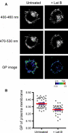
Global condensation of the plasma membrane by the actin cytoskeleton. The lipophilic dye Laurdan is an environment sensitive fluororophore that serves as a reporter of water penetration into membrane bilayers [126]. Increased lipid packing due to lipid ordering results in a blue shift in the Laurdan emission spectrum, from centered at approximately 500 nm in fluid bilayers to 445 nm in ordered membranes [127]. The normalized ratio of emission channels centered on these wavelengths has been used as a measure of relative membrane ordering [128]. T cells were treated with Laurdan before disrupting the actin cytoskeleton by treating with latrunculin B (Lat B). The samples were imaged in two separate channels, represented by emission wavelengths 400–460 and 470–530 nm using a Leica SP2 multi-photon confocal microscope. General polarization (GP), which reflects the relative lipid condensation [128], is therefore an indicator of lipid ordering. GP was calculated for each pixel in the plasma membrane using the equation GP = I(400–460)−I(470–530)/(I(400–460) + I(470–530)) [56]. GP values range between −1.0 and +1.0, and they are directly proportional to the relative membrane condensation. a Images in the indicated channels of untreated control and Lat B-treated T cells. The bottom row shows the calculated GP image for each sample. b Average GP values of the plasma membrane measured in approximately 50 separate untreated or Lat B-treated T cells
Fig. 4
Fig. 4
Actin cytoskeleton-dependent raft organization. a Topography of raft nanoclusters and macrodomains. Rafts occur as nanoclusters that are less than 20 nm in diameter, but can cluster to larger structures to include macrodomains that are microns in size. Both the nanoclusters and larger assembles form in an actin-dependent manner [29, 31, 129]. The macrodomains form as a result of signals that activate actin polymerization and attachment of actin filaments to cell membranes. Examples of raft macrodomains include immunological synapses that form in activated lymphocytes, cell–cell adhesion complexes, leading edge and uropod of migrating cells. b The cortical actin cytoskeleton is tethered to the plasma membrane through protein linkers, and these interactions likely structure rafts in the membrane. The meshwork of cortical actin filaments also compartmentalizes the membrane into areas approximately 50–200 nm in size. The compartments represent areas of transient and nonspecific membrane protein confinement by the underlying actin filaments
Fig. 5
Fig. 5
Model for raft protein regulation by cholesterol and the actin cytoskeleton. The Src family kinase (SFK) Lck is activated by the membrane phosphatase CD45 through dephosphorylation of its regulatory C-terminal tyrosine Tyr505. A significant fraction of Lck associates with rafts, yet CD45 is restricted to the nonraft membrane fraction. Raft association of Lck therefore sequesters it from CD45, leading to reduced activity for the raft-associated pool of molecules. Drug treatments that disrupt rafts, such as filipin and Lat B, eliminate this sequestration, resulting in activation of Lck [29]. Conversely, signals that activate actin polymerization and assembly of rafts, such as that from the TCR in T cells, are predicted to enhance the sequestration and down-regulation of raft-associated Lck

Similar articles

Cited by

References

    1. Rodgers W, Farris D, Mishra S. Merging complexes: properties of membrane raft assembly during lymphocyte signaling. Trends Immunol. 2005;26:97–103. doi: 10.1016/j.it.2004.11.016. - DOI - PubMed
    1. Brown DA, London E. Structure and function of sphingolipid- and cholesterol-rich membrane rafts. J Biol Chem. 2000;275:17221–17224. doi: 10.1074/jbc.R000005200. - DOI - PubMed
    1. Sharma P, Varma R, Sarasij RC, Ira, Gousset K, Krishnamoorthy G, Rao M, Mayor S (2004) Nanoscale organization of multiple GPI-anchored proteins in living cell membranes. Cell 116: 577–589 - PubMed
    1. Zacharias DA, Violin JD, Newton AC, Tsien RY. Partitioning of lipid-modified monomeric GFPs into membrane microdomains of live cells. Science. 2002;296:913–916. doi: 10.1126/science.1068539. - DOI - PubMed
    1. Eggeling C, Ringemann C, Medda R, Schwarzmann G, Sandhoff K, Polyakova S, Belov VN, Hein B, von Middendorff C, Schonle A, Hell SW. Direct observation of the nanoscale dynamics of membrane lipids in a living cell. Nature. 2009;457:1159–1162. doi: 10.1038/nature07596. - DOI - PubMed

Publication types

Substances

LinkOut - more resources