. 2009 May;116(5):587-97.
doi: 10.1007/s00702-009-0226-9. Epub 2009 Apr 16.

Evidence of angiogenic vessels in Alzheimer's disease

Affiliations

Evidence of angiogenic vessels in Alzheimer's disease

Brinda S Desai et al. J Neural Transm (Vienna). 2009 May.

Abstract

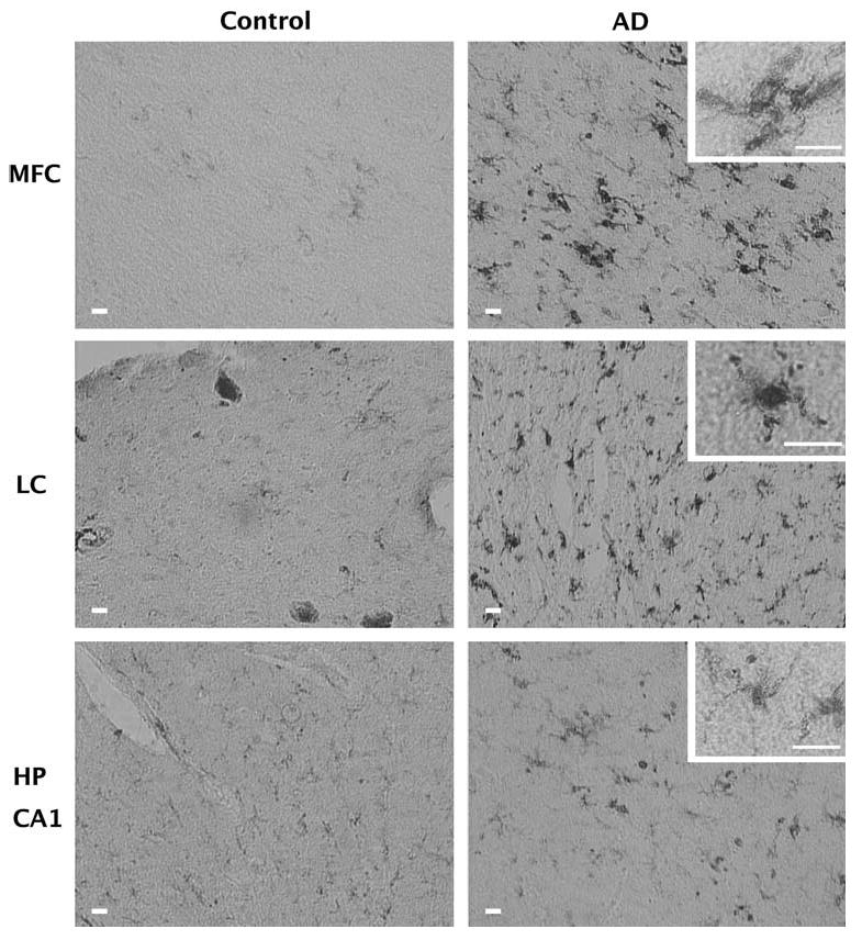
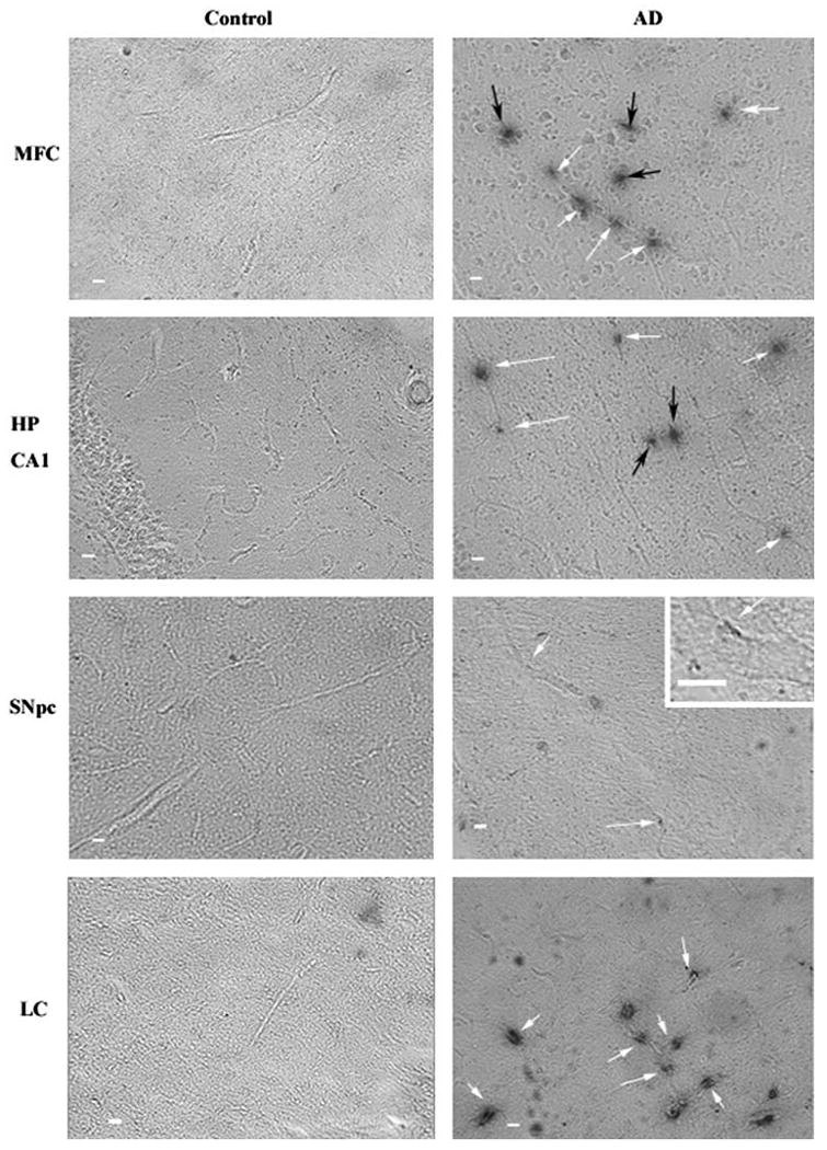
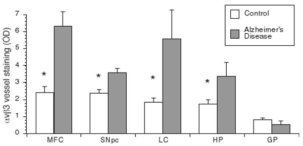
Alterations in the blood brain barrier and brain vasculature may be involved in neurodegeneration and neuroinflammation. We sought to determine if vascular remodeling characterized by angiogenic vessels or increased vascular density, occurred in pathologically confirmed Alzheimer's disease (AD) postmortem human brain tissues. We examined brains of deceased, older catholic clergy from the Religious Order Study, a longitudinal clinical-pathological study of aging and AD. The hippocampus, midfrontal cortex, substantia nigra, globus pallidus and locus ceruleus were examined for integrin alphavbeta3 immunoreactivity, a marker of angiogenesis, and vascular densities. Activated microglia cell counts were also performed. All areas except the globus pallidus exhibited elevated alphavbeta3 immunoreactivity in AD cases compared with controls. Only in the hippocampus did the ongoing angiogenesis result in increased vascular density compared with controls. Vascular density was correlated with Abeta load in the hippocampus and alphavbeta3 reactivity was correlated with neurofibrillary tangles in the midfrontal cortex and in the substantia nigra. These data indicate that ongoing angiogenesis is present in brain regions affected by AD pathology and may be related to tissue injury.


Conflict of interest statement

Conflict of interest statement: There is no actual or potential conflicts of interest.

Figures

Fig. 1
Fig. 1
Activation of microglia in AD. Shown is the immunoreactivity of tissue from the midfrontal cortex (MFC), locus ceruleus (LC), and hippocampus (HP CA1) of control and Alzheimer's subjects to an antibody that recognizes the MHC Class II antigen. This antigen is up regulated on the cell surface of activated microglia. All bars indicate 20 μm. Increased numbers of activated microglia are observed in the MFC, and LC of AD subjects compared with controls. In addition, many microglia from the AD subjects have a morphology (see insets) characteristic of activated microglia. In the Hippocampus, there was no difference between control and AD and there was a greater prevalence of the ramified morphology characteristic of unactivated microglia
Fig. 2
Fig. 2
Intensity of αvβ3 staining in Alzheimer's disease tissue samples. Slices from human postmortem brain tissue were labeled with mouse anti-human integrin αvβ3 antibody and visualized with the chromagen DAB. All images are at ×5 magnification and inset images are ×25 magnification. Scale bar = 10 μm. Arrows indicate αvβ3 positive vessels in control and Alzheimer's disease (AD). Integrin αvβ3 immunoreactivity could be observed in the midfrontal cortex (MFC), hippocampus (HP CA1), SNpc and locus ceruleus (LC) of AD subjects. White arrows indicate αvβ3 staining along a longitudinal vessel. Black arrows indicate staining occurring without an apparent longitudinal vessel. While the black arrows likely indicate cross sectional vessel perpendicular to the plane of the image, they were not counted in the quantitation of vessels in Fig. 3 avoid any false positives due to edge effects around potential holes in the tissue
Fig. 3
Fig. 3
Intensity of αvβ3 vessel staining in postmortem human brain tissue samples. Tissue slices were labeled with αvβ3 as in Fig. 2 and the optical density of longitudinal vessels was measured as described in the methods. Shown is the mean optical density (OD) of vessels for control and AD subjects ± SEM for each brain area. Integrin αvβ3 immunoreactivity is significantly greater in the hippocampus (HP), midfrontal cortex (MFC), SNpc and locus ceruleus (LC) of AD subjects compared with control subjects. There was no difference in αvβ3 staining of vessels between control and AD subjects in the globus pallidus (GP). Data is expressed as Mean ± SEM. *Statistical significance was determined using Student's t tests for mean comparison (p < 0.05)
Fig. 4
Fig. 4
Total number of vessels in postmortem human tissue samples. Vessel numbers were counted under bright field microscopy. Density measurements were obtained by dividing total number of vessels by the area of interest (refer to Methods). Although a change in vessel number was observed in AD subjects, it was only significantly higher in the hippocampus. Data is expressed as Mean ± SEM. *Statistical significance was determined using Student's t tests for mean comparisons (p < 0.05)

References

    1. Abbott NJ. Inflammatory mediators and modulation of blood– brain barrier permeability. Cell Mol Neurobiol. 2000;20:131–147. - PMC - PubMed
    1. Akiyama H, Kawamata T, Dedhar S, McGeer PL. Immunohistochemical localization of vitronectin, its receptor and beta-3 integrin in Alzheimer brain tissue. J Neuroimmunol. 1991;32:19–28. - PubMed
    1. Akiyama H, Ikeda K, Kondo H, McGeer PL. Thrombin accumulation in brains of patients with Alzheimer's disease. Neurosci Lett. 1992;146:152–154. - PubMed
    1. Alzheimer A, Stelzmann RA, Schnitzlein HN, Murtagh FR. An English translation of Alzheimer's 1907 paper, “Uber eine eigenartige Erkankung der Hirnrinde”. Clin Anat. 1995;8:429–431. - PubMed
    1. Bennett DA, Schneider JA, Wilson RS, Bienias JL, Arnold SE. Neurofibrillary tangles mediate the association of amyloid load with clinical Alzheimer disease and level of cognitive function. Arch Neurol. 2004;61:378–384. - PubMed

Publication types

MeSH terms