Comparative Study
. 2009 Jun 9;48(22):4838-45.
doi: 10.1021/bi900166y.

Crystal structure of the human lymphoid tyrosine phosphatase catalytic domain: insights into redox regulation

Affiliations
Comparative Study

Crystal structure of the human lymphoid tyrosine phosphatase catalytic domain: insights into redox regulation

Sophia J Tsai et al. Biochemistry. .

Abstract

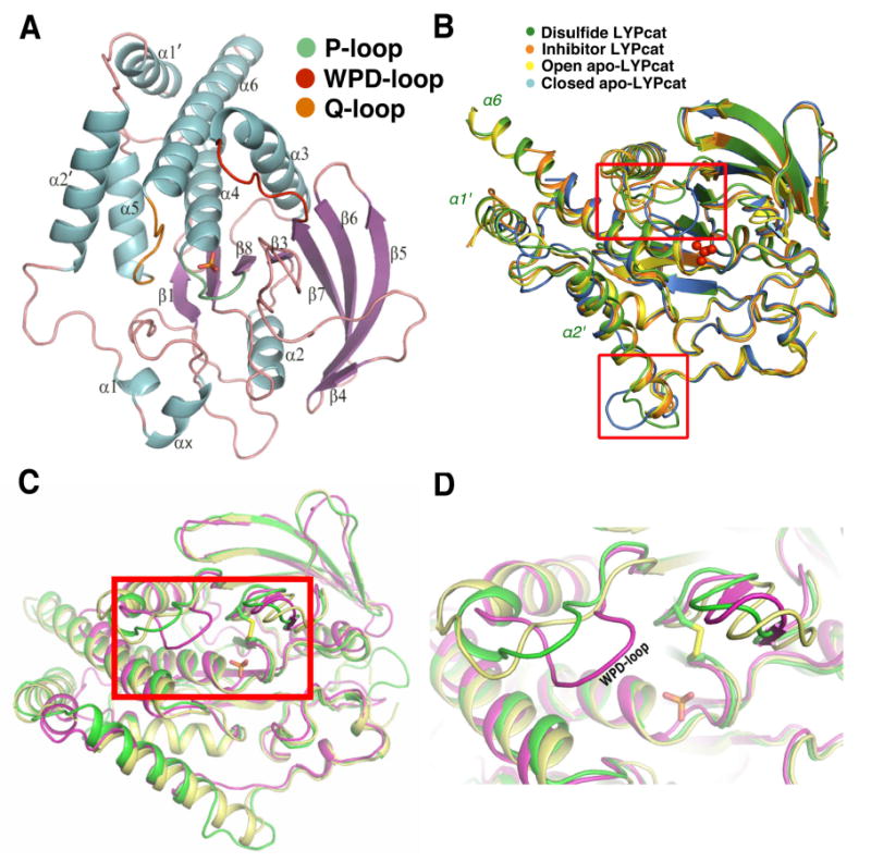
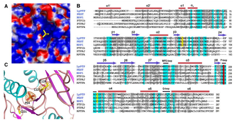
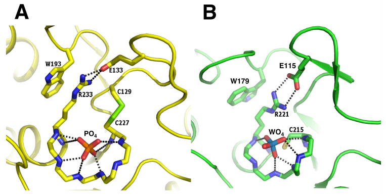
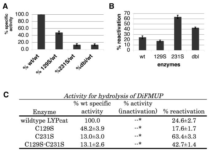
The lymphoid tyrosine phosphatase (LYP), encoded by the PTPN22 gene, recently emerged as an important risk factor and drug target for human autoimmunity. Here we solved the structure of the catalytic domain of LYP, which revealed noticeable differences with previously published structures. The active center with a semi-closed conformation binds a phosphate ion, which may represent an intermediate conformation after dephosphorylation of the substrate but before release of the phosphate product. The structure also revealed an unusual disulfide bond formed between the catalytic Cys and one of the two Cys residues nearby, which is not observed in previously determined structures. Our structural and mutagenesis data suggest that the disulfide bond may play a role in protecting the enzyme from irreversible oxidation. Surprisingly, we found that the two noncatalytic Cys around the active center exert an opposite yin-yang regulation on the catalytic Cys activity. These detailed structural and functional characterizations have provided new insights into autoregulatory mechanisms of LYP function.


Figures

Fig. 1
Fig. 1
LYP Structure and comparison to other PTPs. A. Ribbon diagram of LYP catalytic domain (LTPcat). Alpha helices are in blue, beta sheets are colored purple, the P-loop is colored green, the WPD loop is red, the Q-loop is orange, and all other loops are pink. The bound phosphate ion at the active site is shown as a stick model. B. The superposition of known structures of LYPcat, with the major areas of differences in surface loops indicated by the red boxes. The structure in this report is shown in green, LYPcat with an inhibitor (PDB code: 2QCT) in orange, corresponding structure without inhibitor (PDB code: 2QCJ) in blue, and the LYPcat alone with the PDB code 2P6X in yellow. The phosphate ion is shown in red ball and stick. C. Comparison of the different WPD loop conformations between LYPcat (shown in green), non-phosphate-bound LYPcat (in yellow, PDB code: 2P6X) and the tungstate bound PTP1B (in magenta, PDB code: 2HNQ). Phosphate ion and disulfide bond of LYPcat are shown as sticks. D. Zoomed view of the boxed area in Fig 1C, showing different locations of WPD loop in the three structures.
Fig. 2
Fig. 2
Binding of the phosphate ion in active site of LYPcat and sequence alignment: A. Surface electrostatic potential of phosphate bound LYPcat, phosphate ion (in yellow) is located in a deep cleft of the surface. B. Sequence and secondary structure alignment of LYPcat with other members of the NT4 subfamily and PTP-1B, PTPRJ and RPTPκ. Arrows represent β-strands, bars denote α-helices, and black lines denote catalytically important loops. The catalytic cysteine is highlighted in red and the two other cysteines around the catalytic site are highlighted in yellow. Residues conserved in all the sequences are highlighted in cyan and those showing at least 50% conservation in grey. The names of the NT4 subfamily are given in blue. C. Fo - Fc map is shown around the phosphate ion and disulfide bridge at a contour level of 4.5σ. Map was calculated where these atoms were omitted from the final coordinates. Alpha helices are denoted in cyan, beta sheets are seen in purple, and loops are colored pink.
Fig. 3
Fig. 3
Active site geometries of PTPs. A. Hydrogen bonds made by the active site phosphate ion in LYPcat with the amide nitrogen of the P-loop residues C227, S228, G230, C231, G232 and T234 and the interactions of R233 with W193 and E133. Side-chains of residues 228-232 were omitted for clarity. B. Interactions made by the active site tungstate ion with PTP-1B (coordinates taken from the PDB code 2HNQ), residues corresponding to R233, W193 and E233 are shown for comparison.
Fig. 4
Fig. 4
Phosphatase Activity Assays of LYP Mutants. A. Relative specific activity of each mutant protein compared to wild type using DiFMUP as the substrate. B. Percent activity of the reactivated proteins (H2O2 treated, followed by DTT treatment) relative to its own protein's full activity before H2O2 inactivation. DiFMUP was used as the substrate. All reactivation percentages of the mutants were found to be statistically significantly different from that of the wild type using the paired t-test. C. Summary of the activity assay results using DiFMUP as the substrate. Column 1 lists each protein, column 2 gives the specific activity of each protein, column 3 gives the percent activity of each protein after inactivation by H2O2, column 4 gives the reactivation activity as a percentage of each protein's original activity after 10 minutes of inactivation by H2O2, followed by DTT reactivation for 15 minutes.

References

    1. Hunter T. A thousand and one protein kinases. Cell. 1987;50:823–829. - PubMed
    1. Mustelin T, Abraham RT, Rudd CE, Alonso A, Merlo JJ. Protein tyrosine phosphorylation in T cell signaling. Frontiers in bioscience : a journal and virtual library. 2002;7:d918–969. - PubMed
    1. Mustelin T, Feng GS, Bottini N, Alonso A, Kholod N, Birle D, Merlo J, Huynh H. Protein tyrosine phosphatases. Frontiers in bioscience : a journal and virtual library. 2002;7:d85–142. - PubMed
    1. Fischer EH, Charbonneau H, Tonks NK. Protein tyrosine phosphatases: a diverse family of intracellular and transmembrane enzymes. Science (New York, NY) 1991;253:401–406. - PubMed
    1. Mustelin T, Taskén K. Positive and negative regulation of T-cell activation through kinases and phosphatases. The Biochemical journal. 2003;371:15–27. - PMC - PubMed

Publication types

MeSH terms

Substances

Associated data