

Therapeutically targeting protein-glycan interactions
- PMID: 19371327
- PMCID: PMC2721254
- DOI: 10.1111/j.1476-5381.2009.00226.x
Therapeutically targeting protein-glycan interactions
Abstract
Glycosylation is the most common form of post-translational modifications by which oligosaccharide side chains are covalently attached to specific residues of the core protein. Especially O-linked glycan structures like the glycosaminoglycans were found to contribute significantly to many (patho-)biological processes like inflammation, coagulation, cancer and viral infections. Glycans exert their function by interacting with proteins thereby changing the structure of the interacting proteins and consequently modulating their function. Given the complex nature of cell-surface and extracellular matrix glycan structures, this therapeutic site has been neglected for a long time, the only exception being the antithrombin III-glycan interaction which has been successfully targeted by unfractionated and low-molecular weight heparins for many decades. Due to the recent breakthrough in the '-ome' sciences, among them proteomics and glycomics, protein-glycan interactions became more amenable for therapeutic approaches so that novel inhibitors of this interaction are currently in preclinical and clinical studies. An overview of current approaches, their advantages and disadvantages, is given and the promising potential of pharmacologically interfering with protein-glycan interactions is highlighted here.
Figures
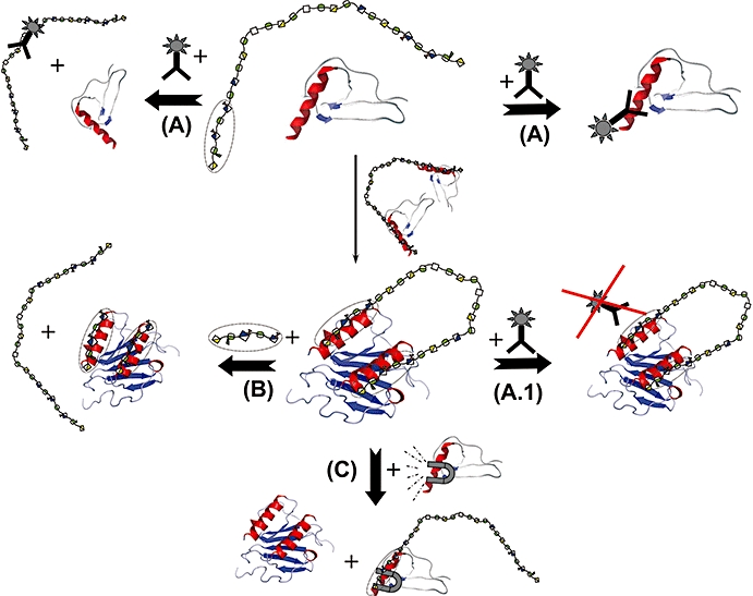
 sequence-specific GAG – Oligo within a proteoglycan side chain;
  antibody. (A) Antibody approach. The binding of a protein to a (specific) GAG chain can be prevented by an antibody to either the (proteo-)glycan or the protein interaction partner. (A.1) The GAG-bound protein may not be targetable by the antibody raised against the soluble protein as structural rearrangements of the protein upon GAG binding as well as the change of overall/surface charge influence/destroy the antibody binding epitope. (B) Glycan- or glycan mimetic-based approach. The protein–GAG interaction can be antagonized by the addition of the synthesized protein-specific GAG sequence (or a mimetic thereof) which displaces the target protein from the natural GAG ligand. (C) Protein-based approach. The protein–GAG interaction can be antagonized by the addition of a mutant form of the target protein which exhibits higher affinity towards the natural GAG ligand and thereby displaces the wild-type target protein from the GAG chain. GAG, glycosaminoglycan.
Similar articles
-
Quantitative O-glycomics based on improvement of the one-pot method for nonreductive O-glycan release and simultaneous stable isotope labeling with 1-(d0/d5)phenyl-3-methyl-5-pyrazolone followed by mass spectrometric analysis.J Proteomics. 2017 Jan 6;150:18-30. doi: 10.1016/j.jprot.2016.08.012. Epub 2016 Aug 29. J Proteomics. 2017. PMID: 27585995
-
Human disease glycomics: technology advances enabling protein glycosylation analysis - part 1.Expert Rev Proteomics. 2018 Feb;15(2):165-182. doi: 10.1080/14789450.2018.1421946. Epub 2018 Jan 11. Expert Rev Proteomics. 2018. PMID: 29285957 Review.
-
Serial in-solution digestion protocol for mass spectrometry-based glycomics and proteomics analysis.Mol Omics. 2020 Aug 1;16(4):364-376. doi: 10.1039/d0mo00019a. Epub 2020 Apr 20. Mol Omics. 2020. PMID: 32309832 Free PMC article.
-
Harnessing glycomics technologies: integrating structure with function for glycan characterization.Electrophoresis. 2012 Mar;33(5):797-814. doi: 10.1002/elps.201100231. Electrophoresis. 2012. PMID: 22522536 Free PMC article. Review.
-
New targets for glycosaminoglycans and glycosaminoglycans as novel targets.Expert Rev Proteomics. 2013 Feb;10(1):77-95. doi: 10.1586/epr.12.75. Expert Rev Proteomics. 2013. PMID: 23414361 Review.
Cited by
-
Heparin/Heparan sulfate proteoglycans glycomic interactome in angiogenesis: biological implications and therapeutical use.Molecules. 2015 Apr 10;20(4):6342-88. doi: 10.3390/molecules20046342. Molecules. 2015. PMID: 25867824 Free PMC article. Review.
-
Structure-based design of decoy chemokines as a way to explore the pharmacological potential of glycosaminoglycans.Br J Pharmacol. 2012 Nov;167(6):1195-205. doi: 10.1111/j.1476-5381.2012.02089.x. Br J Pharmacol. 2012. PMID: 22747966 Free PMC article. Review.
-
Characterizing carbohydrate-protein interactions by nuclear magnetic resonance spectroscopy.Biopolymers. 2013 Oct;99(10):796-806. doi: 10.1002/bip.22329. Biopolymers. 2013. PMID: 23784792 Free PMC article.
-
Designing CXCL8-based decoy proteins with strong anti-inflammatory activity in vivo.Biosci Rep. 2013 Sep 17;33(5):e00068. doi: 10.1042/BSR20130069. Biosci Rep. 2013. PMID: 23919527 Free PMC article.
-
Glycomic profiling of carcinoembryonic antigen isolated from human tumor tissue.Clin Proteomics. 2015 Jun 27;12(1):17. doi: 10.1186/s12014-015-9088-3. eCollection 2015. Clin Proteomics. 2015. PMID: 26157355 Free PMC article.
References
-
- Ashikari S, Habuchi H, Kimata K. Characterization of heparan sulfate oligosaccharides that bind to hepatocyte growth factor. J Biol Chem. 1995;270:29586–29593. - PubMed
-
- Ashikari-Hada S, Habuchi H, Kariya Y, Itoh N, Reddi AH, Kimata K. Characterization of growth factor-binding structures in heparin/heparan sulfate using an octasaccharide library. J Biol Chem. 2004;279:12346–12354. - PubMed
-
- Berg JM, Tymodczko JL, Stryer L. Biochemistry. New York: W. H. Freeman and Company; 2002.
-
- Bishop JR, Schuksz M, Esko JD. Heparan sulphate proteoglycans fine-tune mammalian physiology. Nature. 2007;446:1030–1037. - PubMed
-
- Capila I, Linhardt RJ. Heparin-protein interactions. Angew Chem Int Ed Engl. 2002;41:391–412. - PubMed
Publication types
MeSH terms
Substances
LinkOut - more resources
Full Text Sources
Other Literature Sources
