. 2009 Apr 15;236(2):202-9.
doi: 10.1016/j.taap.2009.01.023. Epub 2009 Feb 6.

Induction of heme oxygenase 1 by arsenite inhibits cytokine-induced monocyte adhesion to human endothelial cells

Affiliations

Induction of heme oxygenase 1 by arsenite inhibits cytokine-induced monocyte adhesion to human endothelial cells

Xi Sun et al. Toxicol Appl Pharmacol. .

Abstract

Heme oxygenase-1 (HO-1) is an oxidative stress responsive gene upregulated by various physiological and exogenous stimuli. Arsenite, as an oxidative stressor, is a potent inducer of HO-1 in human and rodent cells. In this study, we investigated the mechanistic role of arsenite-induced HO-1 in modulating tumor necrosis factor alpha (TNF-alpha) induced monocyte adhesion to human umbilical vein endothelial cells (HUVEC). Arsenite pretreatment, which upregulated HO-1 in a time- and concentration-dependent manner, inhibited TNF-alpha-induced monocyte adhesion to HUVEC and intercellular adhesion molecule 1 protein expression by 50% and 40%, respectively. Importantly, knockdown of HO-1 by small interfering RNA abolished the arsenite-induced inhibitory effects. These results indicate that induction of HO-1 by arsenite inhibits the cytokine-induced monocyte adhesion to HUVEC by suppressing adhesion molecule expression. These findings established an important mechanistic link between the functional monocyte adhesion properties of HUVEC and the induction of HO-1 by arsenite.


Figures
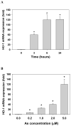
Fig. 1
Fig. 1
Arsenite induces HO-1 expression in HUVEC. The cells were treated with (A) 5μM arsenite for 0–24h, or (B) different arsenite doses for 6h. Total RNA were extracted and reverse transcribed. The expression level of HO-1 was detected by real-time PCR, and normalized as the ratio of density values to the house-keeping β-actin. The mRNA level was presented as relative HO-1 mRNA levels as compared to the control without arsenite treatment. One-way ANOVA with Dunnett’s multiple comparison test indicates *p< 0.05 vs control. Panels are representative of 3 or more experiments. All results were expressed as means±SE.
Fig. 2
Fig. 2
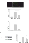
Arsenite attenuates the TNF-α-induced monocyte-endothelial monolayer binding. (A) Fluorescent microscopy of endothelial adhesiveness for monocytes (upper), and quantification of monocyte adhesion to HUVEC (lower); the data were presented by the relative adherent ratio as compared to TNF-α treatment alone. HUVEC were incubated with 5μM arsenite for 6h, followed by addition of 1ng/ml TNF-α for another 6h. HUVEC were then washed with RPMI three times to remove arsenite and TNF-α (to avoid their potential toxicity against the U937 cells); fluorescent labeled U937 cells were added, and incubated at 37°C for 30min. (B) Arsenite suppresses TNF-α-induced ICAM-1 mRNA expression in HUVEC. The cells were incubated with 5μM arsenite for 6h, followed by addition of TNF-α (1ng/ml, 4h). Total RNA were extracted, reverse transcribed, and ICAM-1 mRNA was quantified by real-time PCR; the data were expressed as relative ICAM-1 mRNA levels as compared with untreated control. (C) Arsenite suppresses TNF-α-induced ICAM-1 protein expression in HUVEC. The cells were incubated with 5μM arsenite for 6h, followed by addition of TNF-α (1ng/ml, 4h). Total proteins were then collected after another 14h incubation, 30μg protein was resolved by SDS-PAGE, and ICAM-1 protein level was detected by Western blot (left), and quantitized (right). One-way ANOVA with Turkey’s multiple comparison test indicated significant difference (*p<0.05) between the groups with and without arsenite pre-treatment (panels A–C). Panels are representative of 3 or more experiments. All results were expressed as means±SE.
Fig. 3
Fig. 3
HO-1 siRNA abolishes the inhibitory effect of arsenite on TNF-α-induced monocyte adhesion to HUVEC and ICAM-1 expression. (A) HO-1 siRNA effectively knocks down arsenite-induced HO-1 mRNA expression in HUVEC. The cells transfected with HO-1 siRNA were treated with 5μM arsenite for 6h, and HO-1 mRNA was extracted and quantified. The mRNA data were presented as relative HO-1 mRNA levels compared to the control group of no arsenite/siRNA treatment. Two-way ANOVA with Bonferroni t-test indicates significant difference between the arsenite-treated groups with or without HO-1 siRNA transfection (*p<0.05). (B) HO-1 siRNA effectively knocks down arsenite-induced HO-1 protein expression in HUVEC. The cells transfected with HO-1 siRNA were treated with 5μM arsenite for 20h. HO-1 protein level was analyzed by Western blot. (C) HO-1 siRNA abolishes the inhibitory effect of arsenite on TNF-α-induced monocyte-endothelial monolayer binding. Fluorescent microscopy of endothelial adhesiveness for monocytes (left), and quantification of the monocyte adhesion to HUVEC (right). The data were presented by the relative adherent ratio as compared to the TNF-α/siRNA treated groups. HUVEC cells transfected with HO-1 siRNA were incubated with 5μM arsenite for 6h, followed by addition of 1ng/ml TNF-α for another 6h. The HUVEC cells were washed with RPMI three times (to remove arsenite and TNF-α), the U937 cells were then added to the HUVEC, and incubated at 37°C for 30min. The control is HO-1 siRNA transfected HUVEC cells without TNF-α/arsenite treatment. One-way ANOVA with Turkey’s test indicates significant difference in the TNF-α-treated groups vs control (* p<0.05), but no significant difference between the TNF-α/siRNA and TNF-α+As/siRNA groups. (D) HO-1 siRNA abolishes the suppression of arsenite on TNF-α-induced ICAM-1 protein expression in HUVEC. The cells transfected with HO-1 siRNA were incubated with 5μM arsenite for 6h, followed by addition of 1ng/ml TNF-α. The cells were incubated for another 18h, total proteins were collected, and ICAM-1 was detected by Western blot (left), and quantitized (right). The control is HO-1 siRNA transfected HUVEC cells without TNF-α/arsenite treatment. One-way ANOVA with Turkey’s test indicates * p<0.05 vs control. Panels are representative of 3 or more experiments. All results were expressed as means±SE.

Similar articles

Cited by

References

    1. Andrew AS, Warren AJ, Barchowsky A, Temple KA, Klei L, Soucy NV, O’Hara KA, Hamilton JW. Genomic and proteomic profiling of responses to toxic metals in human lung cells. Environ Health Perspect. 2003;111:825–838. - PMC - PubMed
    1. Balakumar P, Kaur T, Singh M. Potential target sites to modulate vascular endothelial dysfunction: Current perspectives and future directions. Toxicology. 2008;245:49–64. - PubMed
    1. Basu P, Ghosh RN, Grove LE, Klei L, Barchowsky A. Angiogenic potential of 3-nitro-4-hydroxy benzene arsonic acid (Roxarsone) Environ Health Perspect. 2008;116:520–523. - PMC - PubMed
    1. Bau DT, Wang TS, Chung CH, Wang ASS, Jan KY. Oxidative DNA adducts and DNA-protein cross-links are the major DNA lesions induced by arsenite. Environ Health Perspect. 2002;110:753–756. - PMC - PubMed
    1. Benbrahim-Tallaa L, Waterland RA, Styblo M, Achanzar WE, Webber MM, Waalkes MP. Molecular events associated with arsenic-induced malignant transformation of human prostatic epithelial cells: aberrant genomic DNA methylation and K-ras oncogene activation. Toxicol Appl Pharmacol. 2005;206:288–298. - PubMed

Publication types