The cellular context of T cell signaling
- PMID: 19371714
- PMCID: PMC2905632
- DOI: 10.1016/j.immuni.2009.03.010
The cellular context of T cell signaling
Abstract
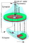
Classical alphabeta T cells protect the host by monitoring intracellular and extracellular proteins in a two-step process. The first step is protein degradation and combination with a major histocompatibility complex (MHC) molecule, leading to surface expression of this amalgam (antigen processing). The second step is the interaction of the T cell receptor with the MHC-peptide complex, leading to signaling in the T cells (antigen recognition). The context for this interaction is a T cell-antigen presenting cell junction, known as an immunological synapse if symmetric and stable and as a kinapse if asymmetric and mobile. The physiological recognition of a ligand takes place most efficiently in the F-actin-rich lamellipodium and is F-actin dependent in stages of formation and triggering and myosin II dependent for signal amplification. This review discusses how these concepts emerged from early studies on adhesion, signaling, and cell biology of T cells.
Figures


References
-
- Aivazian D, Stern LJ. Phosphorylation of T cell receptor zeta is regulated by a lipid dependent folding transition. Nat Struct Biol. 2000;7:1023–1026. - PubMed
-
- Al-Alwan MM, Liwski RS, Haeryfar SM, Baldridge WH, Hoskin DW, Rowden G, West KA. Cutting edge: dendritic cell actin cytoskeletal polarization during immunological synapse formation is highly antigen-dependent. J Immunol. 2003;171:4479–4483. - PubMed
-
- Antón IM, de la Fuente MA, Sims TN, Freeman S, Ramesh N, Hartwig JH, Dustin ML, Geha RS. WIP Deficiency Reveals a Differential Role for WIP and the Actin Cytoskeleton in T and B Cell Activation. Immunity. 2002;16:193–204. - PubMed
-
- Babbitt BP, Allen PM, Matsueda G, Haber E, Unanue ER. Binding of immunogenic peptides to Ia histocompatibility molecules. Nature. 1985;317:359–361. - PubMed
Publication types
MeSH terms
Substances
Grants and funding
LinkOut - more resources
Full Text Sources
Other Literature Sources
Research Materials

