

The cytoskeletal scaffold Shank3 is recruited to pathogen-induced actin rearrangements
- PMID: 19371741
- PMCID: PMC2693461
- DOI: 10.1016/j.yexcr.2009.04.003
The cytoskeletal scaffold Shank3 is recruited to pathogen-induced actin rearrangements
Abstract
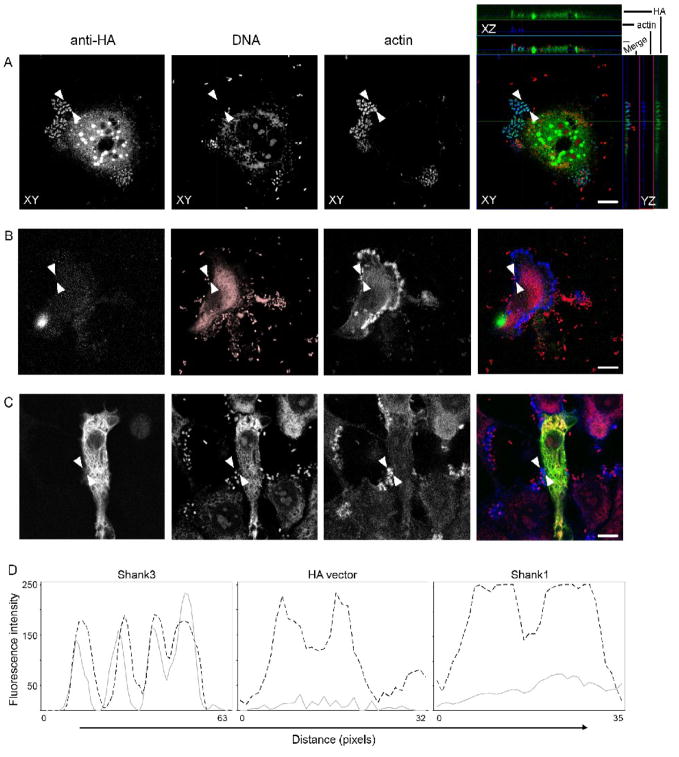
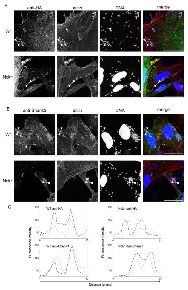
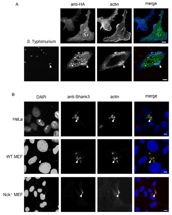
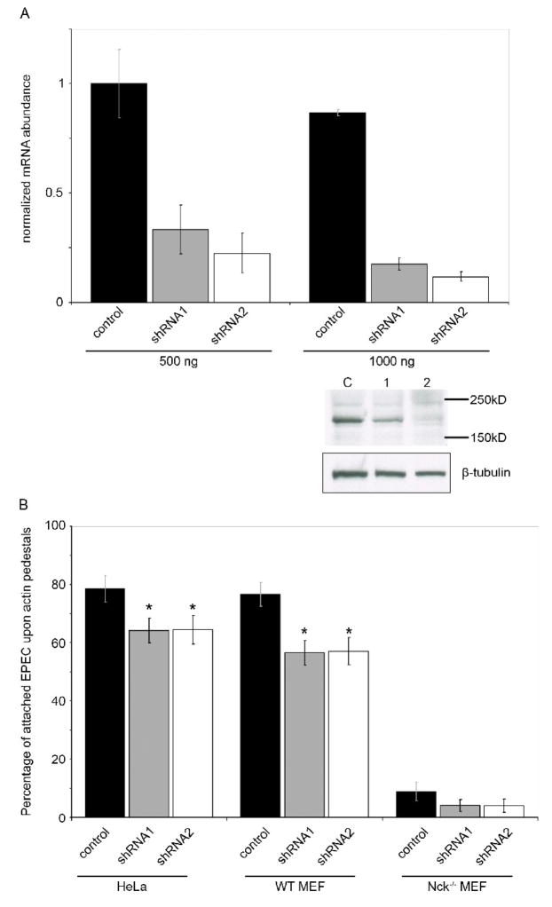
The common gastrointestinal pathogens enteropathogenic Escherichia coli (EPEC) and Salmonella typhimurium both reorganize the gut epithelial cell actin cytoskeleton to mediate pathogenesis, utilizing mimicry of the host signaling apparatus. The PDZ domain-containing protein Shank3, is a large cytoskeletal scaffold protein with known functions in neuronal morphology and synaptic signaling, and is also capable of acting as a scaffolding adaptor during Ret tyrosine kinase signaling in epithelial cells. Using immunofluorescent and functional RNA-interference approaches we show that Shank3 is present in both EPEC- and S. typhimurium-induced actin rearrangements and is required for optimal EPEC pedestal formation. We propose that Shank3 is one of a number of host synaptic proteins likely to play key roles in bacteria-host interactions.
Figures
Similar articles
-
Nck adaptors, besides promoting N-WASP mediated actin-nucleation activity at pedestals, influence the cellular levels of enteropathogenic Escherichia coli Tir effector.Cell Adh Migr. 2014;8(4):404-17. doi: 10.4161/19336918.2014.969993. Cell Adh Migr. 2014. PMID: 25482634 Free PMC article.
-
Enteropathogenic Escherichia coli and vaccinia virus do not require the family of WASP-interacting proteins for pathogen-induced actin assembly.Infect Immun. 2012 Dec;80(12):4071-7. doi: 10.1128/IAI.06148-11. Epub 2012 Sep 10. Infect Immun. 2012. PMID: 22966049 Free PMC article.
-
Role for CD2AP and other endocytosis-associated proteins in enteropathogenic Escherichia coli pedestal formation.Infect Immun. 2010 Aug;78(8):3316-22. doi: 10.1128/IAI.00161-10. Epub 2010 Jun 1. Infect Immun. 2010. PMID: 20515931 Free PMC article.
-
Attaching effacing Escherichia coli and paradigms of Tir-triggered actin polymerization: getting off the pedestal.Cell Microbiol. 2008 Mar;10(3):549-56. doi: 10.1111/j.1462-5822.2007.01103.x. Epub 2007 Dec 4. Cell Microbiol. 2008. PMID: 18053003 Review.
-
Cytoskeletal mechanisms regulating attaching/effacing bacteria interactions with host cells: It takes a village to build the pedestal.Bioessays. 2024 Nov;46(11):e2400160. doi: 10.1002/bies.202400160. Epub 2024 Sep 20. Bioessays. 2024. PMID: 39301984 Review.
Cited by
-
Molecular handoffs in nitrergic neurotransmission.Front Med (Lausanne). 2014 Apr 10;1:8. doi: 10.3389/fmed.2014.00008. eCollection 2014. Front Med (Lausanne). 2014. PMID: 25705621 Free PMC article. Review.
-
Characterization and transplantation of enteric neural crest cells from human induced pluripotent stem cells.Mol Psychiatry. 2018 Mar;23(3):499-508. doi: 10.1038/mp.2016.191. Epub 2016 Oct 25. Mol Psychiatry. 2018. PMID: 27777423 Free PMC article.
-
Altered Intestinal Morphology and Microbiota Composition in the Autism Spectrum Disorders Associated SHANK3 Mouse Model.Int J Mol Sci. 2019 Apr 30;20(9):2134. doi: 10.3390/ijms20092134. Int J Mol Sci. 2019. PMID: 31052177 Free PMC article.
-
Intestinal dysmotility in a zebrafish (Danio rerio) shank3a;shank3b mutant model of autism.Mol Autism. 2019 Jan 31;10:3. doi: 10.1186/s13229-018-0250-4. eCollection 2019. Mol Autism. 2019. PMID: 30733854 Free PMC article.
-
The utility of patient specific induced pluripotent stem cells for the modelling of Autistic Spectrum Disorders.Psychopharmacology (Berl). 2014 Mar;231(6):1079-88. doi: 10.1007/s00213-013-3196-4. Epub 2013 Jul 10. Psychopharmacology (Berl). 2014. PMID: 23839283 Free PMC article. Review.
References
-
- Alto NM, Shao F, Lazar CS, Brost RL, Chua G, Mattoo S, McMahon SA, Ghosh P, Hughes TR, Boone C, Dixon JE. Identification of a bacterial type III effector family with G protein mimicry functions. Cell. 2006;124:133–145. - PubMed
-
- Gonzalez PA, Prado CE, Leiva ED, Carreno LJ, Bueno SM, Riedel CA, Kalergis AM. Respiratory syncytial virus impairs T cell activation by preventing synapse assembly with dendritic cells. Proceedings of the National Academy of Sciences of the United States of America. 2008;105:14999–15004. - PMC - PubMed
Publication types
MeSH terms
Substances
Grants and funding
LinkOut - more resources
Full Text Sources
Molecular Biology Databases
