

A structural approach to skeletal fragility in chronic kidney disease
- PMID: 19371804
- PMCID: PMC2705768
- DOI: 10.1016/j.semnephrol.2009.01.006
A structural approach to skeletal fragility in chronic kidney disease
Abstract
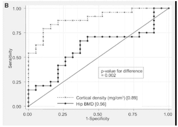
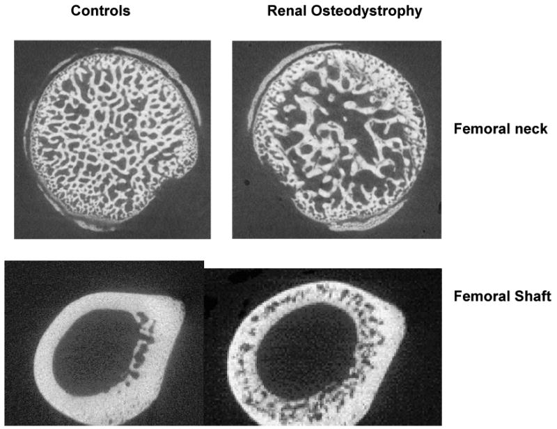
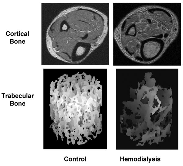
Renal osteodystrophy is a multifactorial disorder of bone metabolism in chronic kidney disease (CKD). As CKD progresses, ensuing abnormalities in mineral metabolism result in distortions in trabecular microarchitecture, thinning of the cortical shell, and increased cortical porosity. Recent studies have shown significantly increased hip fracture rates in CKD stages 3 and 4, in dialysis patients, and in transplant recipients. The majority of studies of bone loss in CKD relied on dual-energy x-ray absorptiometry (DXA) measures of bone mineral density. However, DXA summarizes the total bone mass within the projected bone area, concealing distinct structural alterations in trabecular and cortical bone. Recent data have confirmed that peripheral quantitative computed tomography (pQCT) measures of cortical density and thickness provide substantially better fracture discrimination in dialysis patients, compared with hip or spine DXA. This review summarizes the growing evidence for bone fragility in CKD stages 3 through 5, considers the effects of CKD on trabecular and cortical bone structure as it relates to fracture risk, and details the potential advantages and disadvantages of DXA and alternative measures of bone density, geometry, and microarchitecture, including pQCT, high-resolution pQCT, and micro-magnetic resonance imaging for fracture risk assessment in CKD.
Figures
Similar articles
-
A structural approach to the assessment of fracture risk in children and adolescents with chronic kidney disease.Pediatr Nephrol. 2007 Nov;22(11):1815-24. doi: 10.1007/s00467-007-0490-6. Epub 2007 Jul 11. Pediatr Nephrol. 2007. PMID: 17622566 Free PMC article. Review.
-
Use of dual energy X-ray absorptiometry, the trabecular bone score and quantitative computed tomography in the evaluation of chronic kidney disease-mineral and bone disorders.Nephrology (Carlton). 2017 Mar;22 Suppl 2:19-21. doi: 10.1111/nep.13016. Nephrology (Carlton). 2017. PMID: 28429557 Review.
-
Bone mass and microarchitecture in CKD patients with fracture.J Am Soc Nephrol. 2010 Aug;21(8):1371-80. doi: 10.1681/ASN.2009121208. Epub 2010 Apr 15. J Am Soc Nephrol. 2010. PMID: 20395370 Free PMC article.
-
Skeletal structure in postmenopausal women with osteopenia and fractures is characterized by abnormal trabecular plates and cortical thinning.J Bone Miner Res. 2014;29(5):1101-9. doi: 10.1002/jbmr.2144. J Bone Miner Res. 2014. PMID: 24877245 Free PMC article. Clinical Trial.
-
Spine Trabecular Bone Score as an Indicator of Bone Microarchitecture at the Peripheral Skeleton in Kidney Transplant Recipients.Clin J Am Soc Nephrol. 2017 Apr 3;12(4):644-652. doi: 10.2215/CJN.09850916. Epub 2017 Mar 27. Clin J Am Soc Nephrol. 2017. PMID: 28348031 Free PMC article.
Cited by
-
Age- and gender-related differences in cortical geometry and microstructure: Improved sensitivity by regional analysis.Bone. 2013 Feb;52(2):623-31. doi: 10.1016/j.bone.2012.10.031. Epub 2012 Nov 7. Bone. 2013. PMID: 23142360 Free PMC article.
-
Can one evaluate bone disease in chronic kidney disease without a biopsy?Curr Opin Nephrol Hypertens. 2014 Jul;23(4):431-7. doi: 10.1097/01.mnh.0000447014.36475.58. Curr Opin Nephrol Hypertens. 2014. PMID: 24867672 Free PMC article. Review.
-
Changes in skeletal collagen cross-links and matrix hydration in high- and low-turnover chronic kidney disease.Osteoporos Int. 2015 Mar;26(3):977-85. doi: 10.1007/s00198-014-2978-9. Epub 2014 Dec 3. Osteoporos Int. 2015. PMID: 25466530 Free PMC article.
-
Effects of Cinacalcet on Fracture Events in Patients Receiving Hemodialysis: The EVOLVE Trial.J Am Soc Nephrol. 2015 Jun;26(6):1466-75. doi: 10.1681/ASN.2014040414. Epub 2014 Dec 11. J Am Soc Nephrol. 2015. PMID: 25505257 Free PMC article. Clinical Trial.
-
A comparison of calcium to zoledronic acid for improvement of cortical bone in an animal model of CKD.J Bone Miner Res. 2014 Apr;29(4):902-10. doi: 10.1002/jbmr.2089. J Bone Miner Res. 2014. PMID: 24038306 Free PMC article.
References
-
- Parfitt AM. A structural approach to renal bone disease. J Bone Miner Res. 1998;13(8):1213–20. - PubMed
-
- Alem AM, Sherrard DJ, Gillen DL, Weiss NS, Beresford SA, Heckbert SR, Wong C, Stehman-Breen C. Increased risk of hip fracture among patients with end-stage renal disease. Kidney Int. 2000;58(1):396–9. - PubMed
-
- Stehman-Breen CO, Sherrard DJ, Alem AM, Gillen DL, Heckbert SR, Wong CS, Ball A, Weiss NS. Risk factors for hip fracture among patients with end-stage renal disease. Kidney Int. 2000;58(5):2200–5. - PubMed
-
- Jamal SA, Leiter RE, Jassal V, Hamilton CJ, Bauer DC. Impaired muscle strength is associated with fractures in hemodialysis patients. Osteoporos Int. 2006;17(9):1390–7. - PubMed
-
- Danese MD, Kim J, Doan QV, Dylan M, Griffiths R, Chertow GM. PTH and the risks for hip, vertebral, and pelvic fractures among patients on dialysis. Am J Kidney Dis. 2006;47(1):149–56. - PubMed
Publication types
MeSH terms
Grants and funding
LinkOut - more resources
Full Text Sources
Other Literature Sources
Medical
Research Materials
