. 2009 Jun;77(5):442-9.
doi: 10.1016/j.diff.2009.03.001. Epub 2009 Apr 15.

Mdm4 loss in the intestinal epithelium leads to compartmentalized cell death but no tissue abnormalities

Affiliations

Mdm4 loss in the intestinal epithelium leads to compartmentalized cell death but no tissue abnormalities

Yasmine A Valentin-Vega et al. Differentiation. 2009 Jun.

Abstract

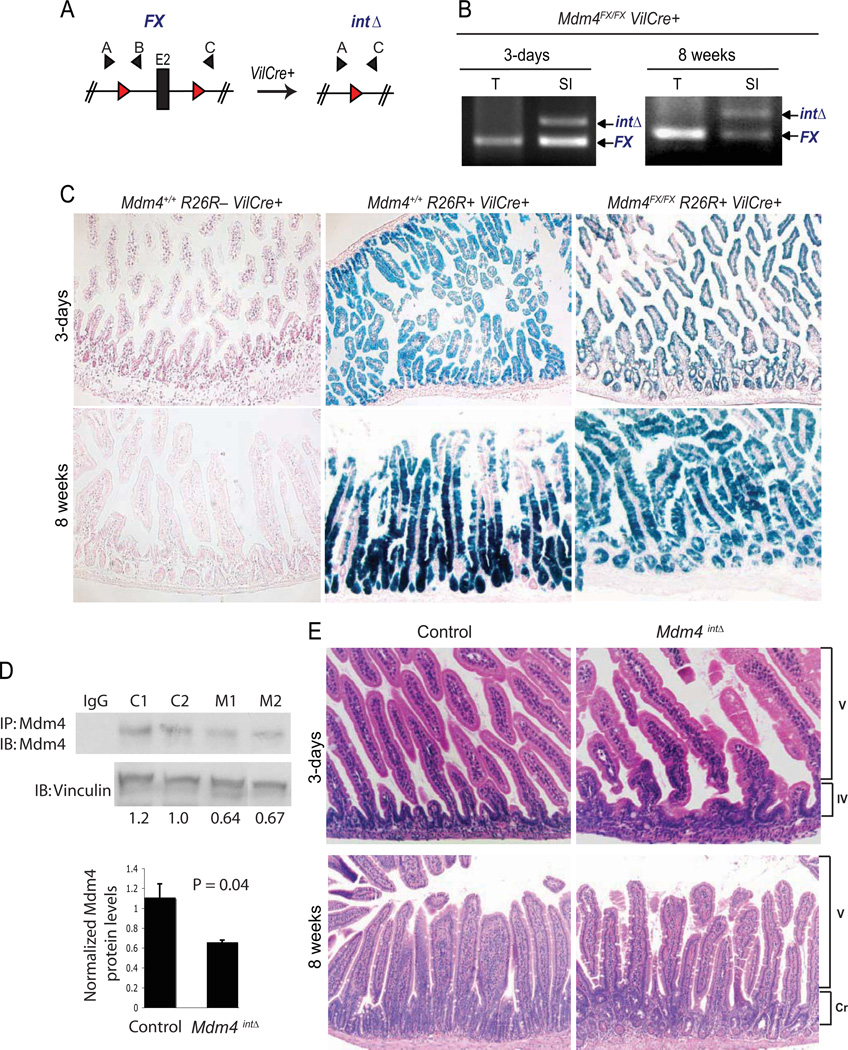
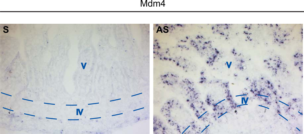
Mdm4 is a critical inhibitor of the p53 tumor suppressor. Mdm4 null mice die early during embryogenesis due to increased p53 activity. In this study, we explore the role that Mdm4 plays in the intestinal epithelium by crossing mice carrying the Mdm4 floxed allele to mice with the Villin Cre transgene. Our data show that loss of Mdm4 (Mdm4intDelta) in this tissue resulted in viable animals with no obvious morphological abnormalities. However, these mutants displayed increased p53 levels and apoptosis exclusively in the proliferative compartment of the intestinal epithelium. This phenotype was completely rescued in a p53 null background. Notably, the observed compartmentalized apoptosis in proliferative intestinal epithelial cells was not due to restricted Mdm4 expression in this region. Thus, in this specific cellular context, p53 is negatively regulated by Mdm4 exclusively in highly proliferative cells.


Figures

Figure 1
Figure 1. Mice survive with loss of Mdm4 in the intestinal epithelium and display normal intestinal morphology
(A) Schematic diagram of the non-recombined (FX) and recombined (intΔ) Mdm4 alleles. The Mdm4 conditional allele targets exon 2 (E2), which contains the translation start site. Red triangles represent loxP sites flanking exon 2; Black triangles represent primer sets used to identify the FX and intΔ alleles (primers A and B amplify the non-recombined (FX) allele while primers A and C amplify the recombined (intΔ) allele). (B) PCR analysis in the intestine of 3-day old Mdm4FX/FX VilCre+ mice using primers A, B, and C. SI, small intestine; T, tail. (C) β-galactosidase activity assays in the small intestine of 3-day and 8-week old mice. Blue denotes cells with β-galactosidase activity (original magnification X100). Mdm4+/+ R26R− VilCre+, negative control; Mdm4+/+ R26R+ VilCre+, positive control; Mdm4FX/FX R26R+ VilCre+, Mdm4intΔ mutant. (D) Mdm4 protein expression in 3-day old whole intestines. Mdm4 protein was immunoprecipitated and then detected by western blot analysis. Mdm4 protein levels were quantified and normalized to the value of Vinculin (loading control). Normalized Mdm4 protein levels were graphed and the p-value was calculated using the Student’s t-test. IgG, Immunoglobulin G (negative control); C1, control #1; C2, control #2; M1, Mdm4intΔ mutant #1; M2, Mdm4intΔ mutant #2. (E) H&E staining of the small intestine of 3-day old pups or intestinal jejunum region of 8-week old mice (original magnification X200 or X100, respectively). V, villus region, IV, intervillus region, Cr, crypt compartment. Intervillus and crypt compartments are regions of undifferentiated cells in young and adult intestines, respectively. The villus compartment contains differentiated intestinal epithelial cells.
Figure 2
Figure 2. Mdm4 regulates p53 specifically in highly proliferative cells of the small intestine
(A) Immunohistochemistry against p53 in the small intestine of 3-day old mice (original magnification X200). Box shows higher magnification of the intervillus (proliferative) region. (B) Caspase-3 immunofluorescence in 3-day old small intestines. Green staining shows caspase-3 positive cells (original magnification X400). (C) TUNEL assay performed in 3-day old small intestines. Black staining illustrates TUNEL positive cells (original magnification X200). (D) Quantification of apoptotic cells in the intervillus pockets of 3-day old small intestines scored by H&E (n = 3 per group). Mdm4intΔ p53−/− genotype represents mice with loss of Mdm4 in the intestine that also lack p53 in the whole body. V, villus (differentiated) region, IV, intervillus (proliferative) region.
Figure 3
Figure 3. Cellular proliferation is intact in the small intestine of Mdm4intΔ mice
(A) Ki-67 immunofluorescence in 3-day old small intestines. Orange fluorescence, cytokeratin (epithelial marker); turquoise fluorescence, Ki-67 (proliferation marker); blue fluorescence, TOPRO-3 (nuclear counterstain); original magnification X400. Graph represents the quantification of the Ki-67 positive staining. (B) Analysis of BrdU incorporation in 8-week old mouse intestines. Mice were injected with 100 µg BrdU/g body weight and sacrificed 2 hr later. BrdU labeled cells (brown) were quantified and values were graphed. The p-value was obtained using Student’s t-test (n = 3 per group); original magnification X200. (C) Reverse transcription RT-PCR analysis of the p53 target, p21. Five mice per group were analyzed and the fold change in p21 mRNA levels was graphed. The p-value was obtained using Student’s t-test.
Figure 4
Figure 4. Mdm4 RNA is expressed in the entire intestinal epithelium of 3-day old mice
In situ hybridization against Mdm4 in 3-day old small intestines of wild-type mice. Black dots represent deposition of Mdm4 RNA in the tissue. S, sense probe; AS, anti-sense probe (original magnification X200). V, villus (differentiated) region, IV, intervillus (proliferative) region.

References

    1. Barak Y, Juven T, Haffner R, Oren M. mdm2 expression is induced by wild type p53 activity. Embo J. 1993;12:461–468. - PMC - PubMed
    1. Boesten LS, Zadelaar SM, De Clercq S, Francoz S, van Nieuwkoop A, et al. Mdm2, but not Mdm4, protects terminally differentiated smooth muscle cells from p53-mediated caspase-3-independent cell death. Cell Death Differ. 2006;13:2089–2098. - PubMed
    1. Chavez-Reyes A, Parant JM, Amelse LL, de Oca Luna RM, Korsmeyer SJ, Lozano G. Switching mechanisms of cell death in mdm2- and mdm4-null mice by deletion of p53 downstream targets. Cancer Res. 2003;63:8664–8669. - PubMed
    1. Evans SC, Viswanathan M, Grier JD, Narayana M, El-Naggar AK, Lozano G. An alternatively spliced HDM2 product increases p53 activity by inhibiting HDM2. Oncogene. 2001;20:4041–4049. - PubMed
    1. Finch RA, Donoviel DB, Potter D, Shi M, Fan A, et al. mdmx is a negative regulator of p53 activity in vivo. Cancer Res. 2002;62:3221–3225. - PubMed

Publication types