. 2009 Jun;296(6):G1370-81.
doi: 10.1152/ajpgi.00074.2009. Epub 2009 Apr 16.

Ano1 is a selective marker of interstitial cells of Cajal in the human and mouse gastrointestinal tract

Affiliations

Ano1 is a selective marker of interstitial cells of Cajal in the human and mouse gastrointestinal tract

Pedro J Gomez-Pinilla et al. Am J Physiol Gastrointest Liver Physiol. 2009 Jun.

Abstract

Populations of interstitial cells of Cajal (ICC) are altered in several gastrointestinal neuromuscular disorders. ICC are identified typically by ultrastructure and expression of Kit (CD117), a protein that is also expressed on mast cells. No other molecular marker currently exists to independently identify ICC. The expression of ANO1 (DOG1, TMEM16A), a Ca(2+)-activated Cl(-) channel, in gastrointestinal stromal tumors suggests it may be useful as an ICC marker. The aims of this study were therefore to determine the distribution of Ano1 immunoreactivity compared with Kit and to establish whether Ano1 is a reliable marker for human and mouse ICC. Expression of Ano1 in human and mouse stomach, small intestine, and colon was investigated by immunofluorescence labeling using antibodies to Ano1 alone and in combination with antibodies to Kit. Colocalization of immunoreactivity was demonstrated by epifluorescence and confocal microscopy. In the muscularis propria, Ano1 immunoreactivity was restricted to cells with the morphology and distribution of ICC. All Ano1-positive cells in the muscularis propria were also Kit positive. Kit-expressing mast cells were not Ano1 positive. Some non-ICC in the mucosa and submucosa of human tissues were Ano1 positive but Kit negative. A few (3.2%) Ano1-positive cells in the human gastric muscularis propria were labeled weakly for Kit. Ano1 labels all classes of ICC and represents a highly specific marker for studying the distribution of ICC in mouse and human tissues with an advantage over Kit since it does not label mast cells.


Figures

Fig. 1.
Fig. 1.
Ano1 immunoreactivity in human gastrointestinal tract. A: labeling patterns for Ano1 immunoreactivity using the mouse (Ms) anti-Ano1 from Applied Genomics (left) and the rabbit (Rb) anti-Ano1 from Abcam (middle) in circular muscle of human stomach (43-yr-old woman). The merged image shows complete colocalization between mouse (red) and rabbit (green in merged images at right) signals. Nuclei were counterstained with DAPI (blue). B: Ano1 immunoreactivity across full thickness of human stomach tissue (33-yr-old woman). MP, myenteric plexus. C: Ano1 immunoreactivity across full thickness of jejunal tissue (41-yr-old man). Arrowhead indicates interstitial cells of Cajal (ICC) in deep muscular plexus (DMP). D: Ano1 immunoreactivity in human colon (40-yr-old woman). Arrowheads indicate submuscular ICC (ICC-SM). A is a single image collected at ×400 magnification. B, C, and D are montages of images collected at ×200 magnification.
Fig. 2.
Fig. 2.
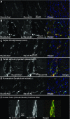
High resolution images of Ano1 immunoreactivity in all subclasses of human ICC. A: Ano1-positive intramuscular ICC (ICC-IM) in circular muscle of the colon (36-yr-old woman). B: positively labeled septal ICC in circular muscle of the jejunum (28-yr-old woman). C: myenteric plexus ICC (ICC-MY) in colon (36-yr-old woman). D: deep muscular plexus ICC (ICC-DMP) in jejunum (28-yr-old woman). E: submuscular ICC (ICC-SM) in colon (40-yr-old woman). F, left: Ano1-positive cells that did not have ICC-like morphology in mucosa from colon (40-yr-old woman). Right: image of the DAPI-labeled nuclei in the same field shown on the left. Dotted line marks nuclei of epithelial cells in a crypt for reference. Scale bar = 20 μm for all panels. All images were collected at ×400 magnification.
Fig. 3.
Fig. 3.
Colocalization of Ano1 and Kit immunoreactivity in the human gastrointestinal tract. In all merged images (right), the signal obtained with rabbit antisera is shown in red and the signal from mouse antisera is shown in green. Blue color shows DAPI staining in nuclei. A: immunoreactivity for Ano1 and Kit were coincident when using mouse anti-Ano1 from Applied Genomics (left) and rabbit anti-Kit from IBL (middle) to label human colonic ICC (40-yr-old woman). B: immunoreactivity for Ano1 and Kit were also coincident when using rabbit anti-Ano1 from Abcam (left) and mouse anti-Kit from Labvision (middle) to label human colonic ICC (53-yr-old man). C: colocalization of Ano1 and Kit immunoreactivity in ICC-IM in the circular muscle from colon (40-yr-old woman). D: colocalization of Ano1 (left) and Kit immunoreactivity (middle) in myenteric and septal ICC of human jejunum (41-yr-old man). E: Ano1 (left) and Kit immunoreactivity (middle) in longitudinal muscle of colon (36-yr-old woman). F: single, confocal slice collected at ×600 magnification in circular muscle of stomach (25-yr-old woman). Ano1 (left) and Kit immunoreactivity (middle) are shown.
Fig. 4.
Fig. 4.
Ano1 labels Kit-negative cells in the mucosa but does not label Kit-positive mast cells. A: in colonic mucosa Ano1 (left, green in merged images at right) and Kit immunoreactivity (middle, red in merged images) does not colocalize as shown in the merged image (right, DAPI stained nuclei in blue). This image is from the same field as shown in Fig. 2F. B: in the muscularis propria, Kit-positive cells (arrowheads, middle, red in merged images) with mast cell morphology do not express Ano1 immunoreactivity (left, green in merged images). Tissue was from stomach (34-yr-old woman). C: mast cell lack of immunoreactivity to Ano1 was confirmed by double labeling human colon sections with antibodies to mast cell tryptase (arrowheads, middle, red in merged images) and Ano1 (left, green in merged images), confirming that mast cells do not express Ano1.
Fig. 5.
Fig. 5.
Presence of weakly Kit-positive (Kit-dim) ICC in the human stomach. A: volume rendered images from confocal stacks collected at ×600 magnification showing Ano1-immunolabeling (left, red in merged images at right) and Kit-immunolabeling (middle, green in merged images) illustrating presence of weakly Kit-positive but Ano1-bright cells with ICC morphology in circular muscle layer of the stomach (25-yr-old woman). Arrowhead points to one of these weakly Kit-positive/Ano1-positive ICC.
Fig. 6.
Fig. 6.
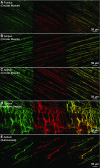
Ano1 antisera label ICC in adult murine stomach. Representative projection images of confocal stacks collected at ×400 magnification from a whole mount incubated with rabbit anti-Ano1 antiserum (Abcam, left, green) and ACK2, a rat anti-Kit antibody (middle, red). The merged images are on the right. ICC-IM are shown from fundus (A), corpus (B), and antrum (C). D: myenteric region ICC in corpus. E: submucosal ICC in antral region.
Fig. 7.
Fig. 7.
Ano1 antisera label ICC in adult murine jejunum and proximal colon. Representative en face projection images of confocal stacks collected at ×400 magnification from a whole mount incubated with rabbit anti-Ano1 antiserum (Abcam, green) and ACK2, a rat anti-Kit antibody (red). A: deep muscular plexus ICC are shown from jejunum. Arrowhead marks an Ano1-positive, Kit-dim ICC. B: myenteric region ICC from jejunum. Asterisk marks a group of Ano1-positive, Kit-dim ICC. C: ICC-IM from circular muscle of proximal colon. D: myenteric region ICC in proximal colon. E: subserosal ICC in proximal colon.
Fig. 8.
Fig. 8.
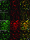
Ano1 antisera label ICC in adult murine distal colon. Representative en face projection images of confocal stacks collected at ×400 magnification from a whole mount incubated with rabbit anti-Ano1 antiserum (Abcam, green in merged images at right) and ACK2, a rat anti-Kit antibody (red). Merged images are on the right. A: submucosal ICC. B: ICC-IM from circular muscle layer. C: myenteric region ICC. D: ICC-IM in longitudinal muscle.

References

    1. Altschul SF, Gish W, Miller W, Myers EW, Lipman DJ. Basic local alignment search tool. J Mol Biol 215: 403–410, 1990. - PubMed
    1. Barajas-Lopez C, Berezin I, Daniel EE, Huizinga JD. Pacemaker activity recorded in interstitial cells of Cajal of the gastrointestinal tract. Am J Physiol Cell Physiol 257: C830–C835, 1989. - PubMed
    1. Berezin I, Huizinga JD, Daniel EE. Interstitial cells of Cajal in the canine colon: a special communication network at the inner border of the circular muscle. J Comp Neurol 273: 42–51, 1988. - PubMed
    1. Burns AJ, Lomax AE, Torihashi S, Sanders KM, Ward SM. Interstitial cells of Cajal mediate inhibitory neurotransmission in the stomach. Proc Natl Acad Sci USA 93: 12008–120013, 1996. - PMC - PubMed
    1. Caputo A, Caci E, Ferrera L, Pedemonte N, Barsanti C, Sondo E, Pfeffer U, Ravazzolo R, Zegarra-Moran O, Galietta LJ. TMEM16A, a membrane protein associated with calcium-dependent chloride channel activity. Science 322: 590–594, 2008. - PubMed

Publication types

MeSH terms

LinkOut - more resources