. 2009 Jul;202(1):77-86.
doi: 10.1677/JOE-09-0119. Epub 2009 Apr 16.

The role of calcium influx pathways in phospholipase D activation in bovine adrenal glomerulosa cells

Affiliations

The role of calcium influx pathways in phospholipase D activation in bovine adrenal glomerulosa cells

Haixia Qin et al. J Endocrinol. 2009 Jul.

Abstract

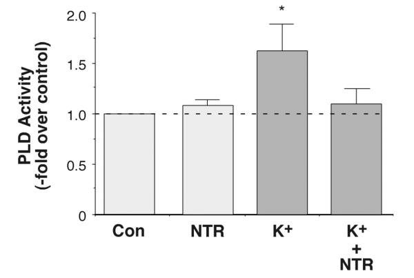
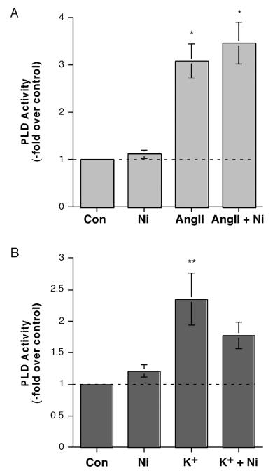
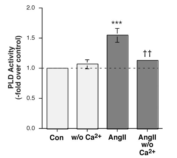
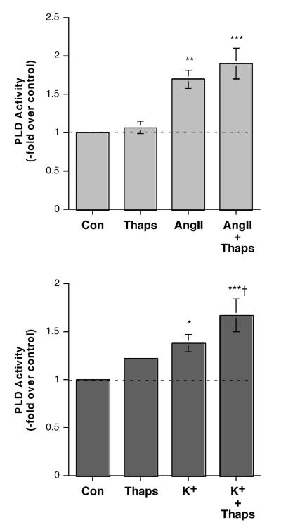
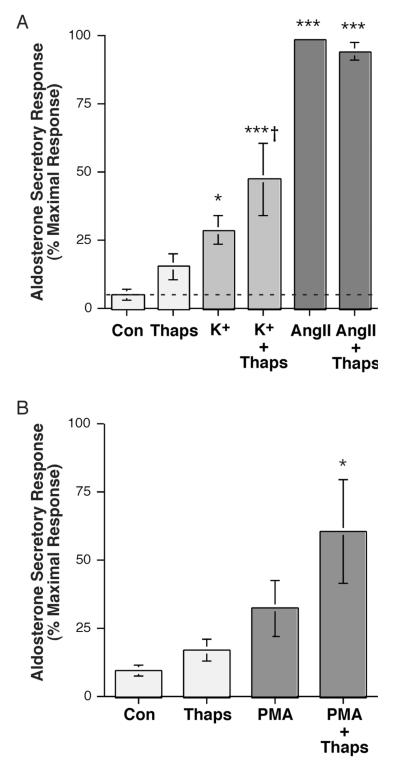
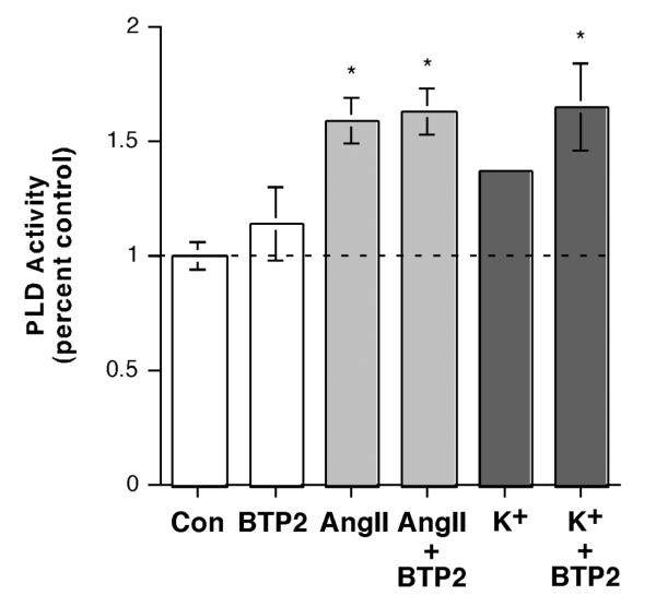
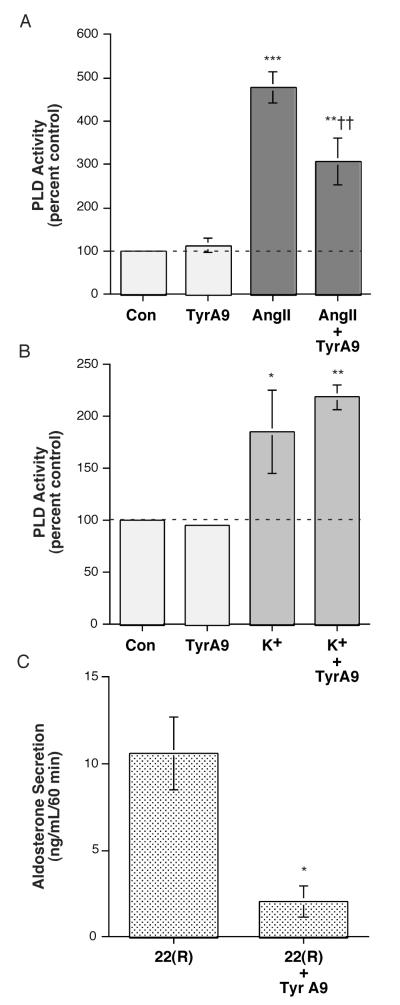
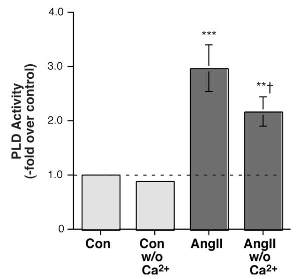
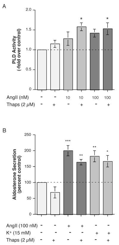
The steroid hormone aldosterone maintains sodium homeostasis and is therefore important in the control of blood volume and pressure. Angiotensin II (AngII) and elevated extracellular potassium concentrations ([K(+)](e)), the prime physiologic regulators of aldosterone secretion from adrenal glomerulosa cells, activate phospholipase D (PLD) in these cells. The role of Ca(2+) in the activation by these agents is unknown, although nitrendipine, a voltage-dependent Ca(2+) channel antagonist, does not inhibit AngII-elicited PLD activation, despite the fact that this compound blocked elevated [K(+)](e)-stimulated PLD activity. PLD activation triggered by AngII was also unaffected by the T-type calcium channel inhibitor nickel. Nevertheless, Ca(2+) influx was required for AngII-induced PLD activation in both primary cultures of bovine adrenal glomerulosa cells and a glomerulosa cell model, the NCI H295R adrenocortical carcinoma cell line. The involvement of store-operated Ca(2+) (SOC) influx and Ca(2+) release-activated Ca(2+) (CRAC) influx pathways in PLD activation was investigated using thapsigargin, an endoplasmic reticulum Ca(2+) pump inhibitor that empties the store to induce SOC influx, and the SOC inhibitor YM-58483 (BTP2), as well as a CRAC inhibitor, tyrphostin A9. In bovine glomerulosa cells, tyrphostin A9 inhibited AngII-induced PLD activation without affecting elevated [K(+)](e)-stimulated enzyme activity. On the other hand, differences were observed between the bovine adrenal glomerulosa and H295R cells in the involvement of Ca(2+) influx pathways in PLD activation, with the involvement of the SOC pathway suggested in the H295R cells. In summary, our results indicate that Ca(2+) entry only through certain Ca(2+) influx pathways is linked to PLD activation.


Figures

Figure 1
Figure 1
Inhibition of Ca2+ Influx with Nitrendipine Completely Inhibited Elevated [K+]e-elicited PLD Activation. [3H]Oleate-prelabeled cells were incubated for 30 minutes with KRB+ containing 3.5 mM KCl (3.5 mM K+-KRB+, control), 1 μM nitrendipine (NTR) in 3.5 mM K+-KRB+, 15 mM K+-KRB+ or 1 μM NTR in 15 mM K+-KRB+ in the presence of 0.5% ethanol. [Note that NaCl in the medium was substituted for by KCl to maintain osmolarity in the 15 mM K+-KRB+ as in (Betancourt-Calle et al., 2001).] Reactions were terminated by the addition of 0.2% SDS containing 5 mM EDTA, and [3H]phosphatidylethanol was extracted, separated by thin-layer chromatography and quantified as in (Jung et al., 1998). Values are expressed as -fold over the control and represent the means (± SEM) of 5 separate experiments; *p<0.05 versus the control value.
Figure 2
Figure 2
Inhibition of alpha1H T-type Ca2+ Channels with Nickel Inhibited Elevated [K+]e-, but Not AngII-, elicited PLD Activation. (A) [3H]Oleate-prelabeled cells were incubated for 30 minutes with KRB+ containing 0.5% ethanol and with and without 10 nM AngII in the presence or absence of 50 μM nickel (Ni) for 30 minutes. Reactions were terminated by the addition of 0.2% SDS containing 5 mM EDTA, and [3H]phosphatidylethanol was extracted, separated by thin-layer chromatography and quantified as in (Jung et al., 1998). Values are expressed as -fold over the control and represent the means (± SEM) of 4 separate experiments; *p<0.05 versus the control value. (B) [3H]Oleate-prelabeled cells were incubated for 30 minutes with 3.5 mM K+-KRB+ or 15 mM K+-KRB+ containing 0.5% ethanol in the absence or presence of 50 μM nickel (Ni) for 30 minutes, and PLD activity monitored as above. Values are expressed as -fold over the control and represent the means (± S.E.M.) of 4 separate experiments; **p<0.01 versus the control value.
Figure 3
Figure 3
AngII-induced PLD Activation was Modulated by Ca2+ in Primary Bovine Adrenal Glomerulosa Cells. Bovine adrenal glomerulosa cells were prelabeled for 20-24 hours with 2.5-5 μCi/mL [3H]oleate in serum-free medium. Cells were then stimulated with or without 10 nM AngII in the presence of 0.5% ethanol in medium containing 1.2 mM Ca2+ or no added Ca2+ for 30 minutes. Lipids were extracted and separated by thin-layer chromatography. PLD was measured as the radioactivity in phosphatidylethanol (PEt) relative to the control, and values are expressed as the means ± SEM of 4 separate experiments; ***p<0.001 versus the control value; ††p<0.01 versus the value in the presence of Ca2+.
Figure 4
Figure 4
Thapsigargin Had Little or No Effect on AngII-Induced PLD Activation, but Enhanced the Effect of Elevated [K+]e on PLD Activity in Bovine Adrenal Glomerulosa Cells. (A) Bovine adrenal glomerulosa cells were prelabeled for 20-24 hours with 2.5-5 μCi/mL [3H]oleate in serum-free medium. Cells were then stimulated with or without 10 nM AngII in the presence or absence of 2 μM thapsigargin (Thaps) or vehicle (0.1-0.2% DMSO) in the presence of 0.5% ethanol for 30 minutes. Lipids were extracted and separated by thin-layer chromatography. Values represent the radioactivity in phosphatidylethanol relative to the control and are expressed as the means ± S.E.M. of 9 separate experiments; *p<0.05 versus the control value. (B) [3H]Oleate-prelabeled bovine adrenal glomerulosa cells were incubated with 3.5 mM K+-KRB+ or 15 mM K+-KRB+ in the presence or absence of 2 μM thapsigargin (Thaps) or vehicle (0.1-0.2% DMSO) in the presence of 0.5% ethanol for 30 minutes. Lipids were extracted and separated by thin-layer chromatography. Values represent the radioactivity in phosphatidylethanol relative to the control and are expressed as the means ± S.E.M. of 5 separate experiments; **p<0.01 versus the control value; †p<0.05 versus K+ alone.
Figure 5
Figure 5
Thapsigargin Had Little or No Effect Alone or on AngII-Stimulated Aldosterone Secretion but Enhanced Steroidogenesis in Response to Elevated [K+]e and PMA in Primary Cultures of Bovine Adrenal Glomerulosa Cells. (A) Bovine adrenal glomerulosa cells were incubated for 1 hour with a bicarbonate-buffered Kreb’s Ringer solution with 2.5 mM sodium acetate (KRB+) containing no additions (Con), 15 mM KCl (K+; iso-osmotic substitution with Na+) or 10 nM AngII in the presence or absence of 2 μM thapsigargin (Thaps) or vehicle (0.1-0.2% DMSO). Supernatants were collected and assayed for aldosterone secretion by radioimmunoassay. Values represent the means ± S.E.M. of 4-5 separate experiments; *p<0.05, **p<0.01 versus control by ANOVA and a Student-Newmann-Keuls post-hoc test. (B) Bovine adrenal glomerulosa cells were incubated for 1 hour with a bicarbonate-buffered Kreb’s Ringer solution with 2.5 mM sodium acetate (KRB+) containing no additions (Con) or 100 nM PMA in the presence or absence of 2 μM thapsigargin (Thaps) or vehicle (0.1-0.2% DMSO). Supernatants were collected and assayed for aldosterone secretion by radioimmunoassay. Values represent the means ± S.E.M. of 4-5 separate experiments, expressed as the percentage of the maximal aldosterone secretory response; *p<0.05, **p<0.01 versus control by ANOVA and a Student-Newmann-Keuls post-hoc test.
Figure 6
Figure 6
Inhibition of Store-operated Ca2+ Influx with BTP2 (YM-58483) Had No Effect on AngII-induced, and Increased Elevated [K+]e-elicited PLD Activation. (A) [3H]Oleate-prelabeled cells were incubated for 30 minutes with KRB+ containing 0.5% ethanol and vehicle (0.1% DMSO) or 10 nM AngII in the presence or absence of 1 μM BTP2 for 30 minutes. Reactions were terminated by the addition of 0.2% SDS containing 5 mM EDTA, and [3H]phosphatidylethanol was extracted, separated by thin-layer chromatography and quantified as in (Jung et al., 1998). Values are expressed as -fold over the control and represent the means (± SEM) of 4 separate experiments; *p<0.05 versus the control value. (B) [3H]Oleate-prelabeled cells were incubated for 30 minutes with 3.5 mM K+-KRB+ or 15 mM K+-KRB+ containing 0.5% ethanol with vehicle (0.1% DMSO) or 1 μM BTP2 for 30 minutes, and PLD activity monitored as above. Values are expressed as -fold over the control and represent the means (± S.E.M.) of 4 separate experiments; *p<0.05 versus the control value.
Figure 7
Figure 7
Inhibition of Ca2+ Release-activated Ca2+ Influx with Tyrphostin A9 Inhibited AngII-, but not Elevated [K+]e-, elicited PLD Activation. (A) [3H]Oleate-prelabeled cells were incubated for 30 minutes with KRB+ containing 0.5% ethanol and vehicle (0.1% DMSO, control) or 10 nM AngII (+ 0.1% DMSO) in the presence or absence of 10 μM tyrphostin A9 (TA9) for 30 minutes. Reactions were terminated by the addition of 0.2% SDS containing 5 mM EDTA, and [ 3H]phosphatidylethanol was extracted, separated by thin-layer chromatography and quantified as in (Jung et al., 1998). Values are expressed as -fold over the control and represent the means (± SEM) of 5 separate experiments; **p<0.01, ***p<0.001 versus the control value, ††p<0.01 versus AngII alone. (B) [3H]Oleate-prelabeled cells were incubated for 30 minutes with 3.5 mM K+-KRB+ or 15 mM K+-KRB+ containing 0.5% ethanol and vehicle (0.1% DMSO, control) or 10 μM tyrphostin A9 (TA9) for 30 minutes, and PLD activity monitored as above. Values are expressed as -fold over the control and represent the means (± S.E.M.) of 4 separate experiments; *p<0.05, **p<0.01 versus the control value. (C) Bovine adrenal glomerulosa cells were incubated for 1 hour with KRB+ containing no additions (Con) or 10 μM 22(R)-hydroxycholesterol in the presence or absence of 10 μM tyrphostin A9 (TA9) or vehicle (0.1-0.2% DMSO). Supernatants were collected and assayed for aldosterone secretion by radioimmunoassay. Values represent the means ± S.E.M., expressed as the ng aldosterone/mL/60 minutes, of 3 separate experiments; *p<0.03 versus 22(R)-hydroxycholesterol alone, as determined using an unpaired Student’s t-test. Note that 22(R)-hydroxycholesterol induced a significant, approximately 11,000-fold increase over the control aldosterone secretory value.
Figure 8
Figure 8
AngII-induced PLD Activation was Modulated by Ca2+ in NCI H295R Adrenocortical Carcinoma Cells. H295R cells were prelabeled for 20-24 hours with 2.5-5 μCi/mL [3H]oleate in serum-free medium. Cells were then stimulated with or without 100 nM AngII in KRB+ containing 1.2 mM Ca2+ or no added Ca2+ in the presence of 0.5% ethanol for 5 minutes. Lipids were extracted and separated by thin-layer chromatography. Values represent the radioactivity in phosphatidylethanol (PEt) relative to the control and are expressed as the means ± S.E.M. of 4 separate experiments; **p<0.01, ***p<0.01 versus the control value, †p<0.05 versus the value in the presence of Ca2+.
Figure 9
Figure 9
Thapsigargin Enhanced the Effect of AngII on PLD Activation Cells but Had No Effect on AngII- (or Elevated [K+]e-) Stimulated Aldosterone Secretion in H295R. (A) H295R cells were pre-labeled for 20-24 hours with 2.5-5 μCi/mL [3H]oleate in serum-free medium. Cells were then stimulated with or without 10 or 100 nM AngII in the presence or absence of 2 μM thapsigargin (Thaps) or vehicle (0.2% DMSO) in the presence of 0.5% ethanol. Lipids were extracted and separated by thin-layer chromatography. Values represent the radioactivity in phosphatidylethanol relative to the control and are expressed as the means ± S.E.M. of 3 separate experiments; *p<0.05 versus the control value. (B) H295R cells were incubated for 5 hours with medium containing no additions (Con), 15 mM KCl (K+) or 100 nM AngII in the presence or absence of 2 μM thapsigargin (Thaps) or vehicle (0.1-0.2% DMSO) as indicated. Supernatants were collected and assayed for aldosterone secretion by radioimmunoassay. Values represent the means ± S.E.M. of 4-5 separate experiments; *p<0.05, **p<0.01, ***p<0.001 versus the control value.

Similar articles

Cited by

References

    1. Aptel HB, Burnay MM, Rossier MF, Capponi AM. The role of tyrosine kinases in capacitative calcium influx-mediated aldosterone production in bovine adrenal zona glomerulosa cells. J Endocrinol. 1999;163(1):131–138. - PubMed
    1. Barrett PQ, Bollag WB, Isales CM, McCarthy RT, Rasmussen H. Role of calcium in angiotensin II-mediated aldosterone secretion. Endocr Rev. 1989;10(4):1–22. - PubMed
    1. Barrett PQ, Ertel EA, Smith MM, Nee JJ, Cohen CJ. Voltage-gated calcium currents have two opposing effects on the secretion of aldosterone. Am. J. Physiol. 1995;268:C985–C992. - PubMed
    1. Betancourt-Calle S, Jung EM, White S, Calle RA, Rasmussen H, Bollag WB. Elevated K + induces MARCKS phosphorylation and phospholipase D activation in glomerulosa cells. Mol. Cell. Endocrinol. 2001;184:65–76. - PubMed
    1. Bird IM, Hanley NA, Word RA, Mathis JM, McCarthy JL, Mason JI, Rainey WE. Human NCI-H295 adrenocortical carcinoma cells: A model for angiotensin-II-responsive aldosterone secretion. Endocrinology. 1993;133:1555–1561. - PubMed

Publication types

MeSH terms