. 2009 Jul;76(1):47-57.
doi: 10.1124/mol.109.055376. Epub 2009 Apr 16.

Thiazolidinediones mimic glucose starvation in facilitating Sp1 degradation through the up-regulation of beta-transducin repeat-containing protein

Affiliations

Thiazolidinediones mimic glucose starvation in facilitating Sp1 degradation through the up-regulation of beta-transducin repeat-containing protein

Shuo Wei et al. Mol Pharmacol. 2009 Jul.

Abstract

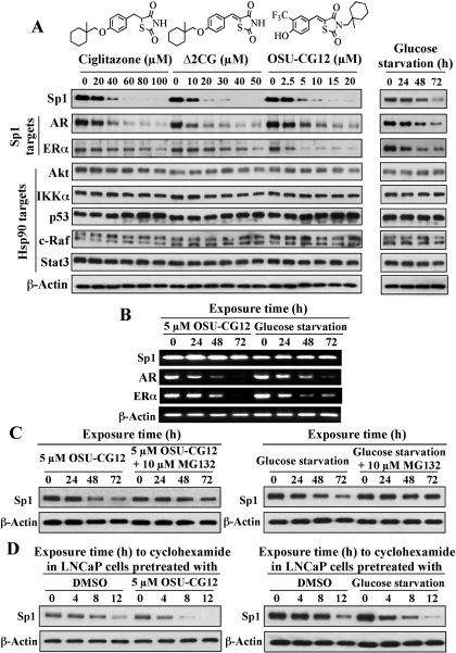
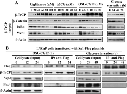
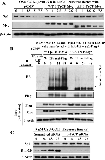
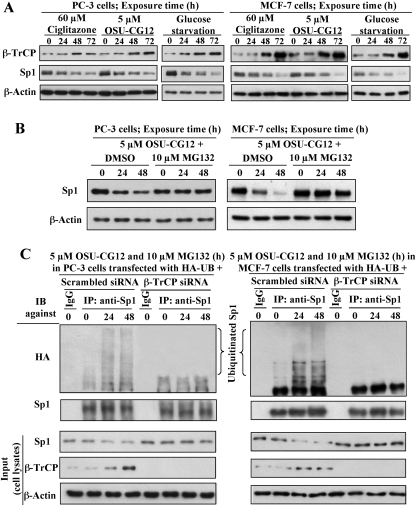
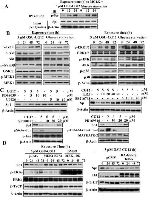
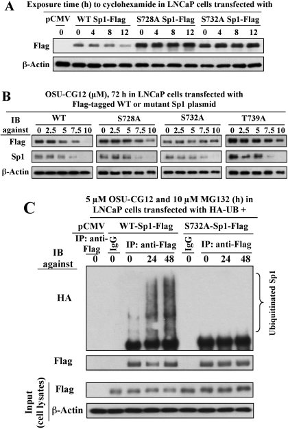
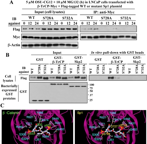
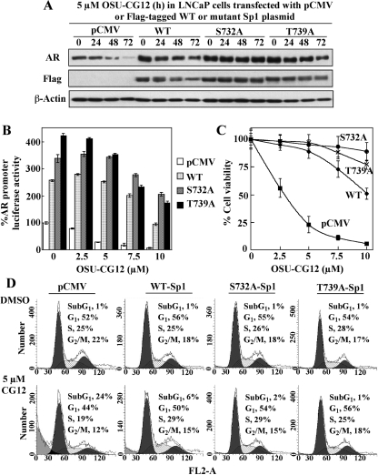
This study investigated the mechanism by which the transcription factor Sp1 is degraded in prostate cancer cells. We recently developed a thiazolidinedione derivative, (Z)-5-(4-hydroxy-3-trifluoromethylbenzylidene)-3-(1-methylcyclohexyl)-thiazolidine-2,4-dione (OSU-CG12), that induces Sp1 degradation in a manner paralleling that of glucose starvation. Based on our finding that thiazolidinediones suppress beta-catenin and cyclin D1 by up-regulating the E3 ligase SCF(beta-TrCP), we hypothesized that beta-transducin repeat-containing protein (beta-TrCP) targets Sp1 for proteasomal degradation in response to glucose starvation or OSU-CG12. Here we show that either treatment of LNCaP cells increased specific binding of Sp1 with beta-TrCP. This direct binding was confirmed by in vitro pull-down analysis with bacterially expressed beta-TrCP. Although ectopic expression of beta-TrCP enhanced the ability of OSU-CG12 to facilitate Sp1 degradation, suppression of endogenous beta-TrCP function by a dominant-negative mutant or small interfering RNA-mediated knockdown blocked OSU-CG12-facilitated Sp1 ubiquitination and/or degradation. Sp1 contains a C-terminal conventional DSG destruction box ((727)DSGAGS(732)) that mediates beta-TrCP recognition and encompasses a glycogen synthase kinase 3beta (GSK3beta) phosphorylation motif (SXXXS). Pharmacological and molecular genetic approaches and mutational analyses indicate that extracellular signal-regulated kinase-mediated phosphorylation of Thr739 and GSK3beta-mediated phosphorylation of Ser728 and Ser732 were critical for Sp1 degradation. The ability of OSU-CG12 to mimic glucose starvation to activate beta-TrCP-mediated Sp1 degradation has translational potential to foster novel strategies for cancer therapy.


Figures

Fig. 1.
Fig. 1.
Evidence that the ability of ciglitazone, Δ2CG, and OSU-CG12 to facilitate the proteasomal degradation of Sp1 in LNCaP cells parallels that of glucose starvation. A, chemical structures of ciglitazone, Δ2CG, and OSU-CG12, and Western blot analysis of the effects of individual thiazolidinediones and glucose starvation on the expression of Sp1 and its downstream targets, AR and ERα, and the Hsp90 client proteins Akt, IKKα, p53, c-Raf, and Stat3. Cells were treated with thiazolidinediones for 72 h or with glucose-free medium. Cell lysates were immunoblotted for the indicated proteins. B, reverse transcription-polymerase chain reaction analysis of the time-dependent effect of 5 μM OSU-CG12 versus glucose starvation on the mRNA levels of Sp1, AR, and ERα. C, the proteasome inhibitor MG132 (10 μM) rescued Sp1 from degradation after treatment of LNCaP cells with 5 μM OSU-CG12 (left) or glucose starvation (right). Cells were treated for the indicated times with OSU-CG12 or glucose starvation alone and in combination with MG132. Cell lysates were immunoblotted for the indicated proteins. D, OSU-CG12 (left) and glucose starvation (right) shortened the half-life of Sp1 in LNCaP cells. Cells were treated with DMSO, 5 μM OSU-CG12, or glucose-deprived RPMI 1640 medium for 12 h, followed by exposure to 100 μg/ml cycloheximide for the indicated time intervals. Cell lysates were immunoblotted with anti-Sp1 and anti-β-actin antibodies.
Fig. 2.
Fig. 2.
A mechanistic link between β-TrCP up-regulation and Sp1 degradation in response to thiazolidinedione treatment or glucose starvation. A, Western blot analysis of the dose-dependent effect of ciglitazone, Δ2CG, and OSU-CG12 vis-à-vis the time-dependent effect of glucose starvation on modulating the expression levels of β-TrCP and its substrates including β-catenin, IκBα, and Wee1 in LNCaP cells. Cells were treated with thiazolidinediones for 72 h or with glucose-free medium. Cell lysates were immunoblotted for the indicated proteins. B, OSU-CG12 and glucose starvation promote the association of Sp1 with the F-box protein β-TrCP. LNCaP cells transiently transfected with the Sp1-Flag plasmid were exposed to 5 μM OSU-CG12 for 6 or 18 h or glucose starvation for 18 or 42 h followed by cotreatment with the proteasome inhibitor MG132 for an additional 6 h. The cell lysates were immunoblotted with antibodies against Flag or various F-box proteins (input), or immunoprecipitated with anti-Flag antibody-agarose conjugates. Equal amounts of the immunoprecipitated proteins were immunoblotted for the indicated proteins.
Fig. 3.
Fig. 3.
Evidence that β-TrCP is involved in OSU-CG12-mediated Sp1 degradation in LNCaP cells. A, effects of ectopically expressed WT β-TrCP-Myc and ΔF-β-TrCP-Myc on OSU-CG12-mediated Sp1 proteolysis. LNCaP cells transiently transfected with plasmids encoding the Myc-tagged WT β-TrCP or ΔF-β-TrCP proteins, or the empty vector as control, were treated with OSU-CG12 as indicated. Cell lysates were immunoblotted with anti-Sp1 and anti-Myc antibodies. B, ectopic expression of ΔF-β-TrCP-Myc blocked OSU-CG12-mediated Sp1 ubiquitination. LNCaP cells cotransfected with plasmids encoding HA-ubiquitin, Sp1-Flag, and WT- or ΔF-β-TrCP-Myc were treated with 5 μM OSU-CG12 for 12 or 36 h, followed by cotreatment with proteasome inhibitor MG132 (10 μM) for an additional 12 h. Equal amounts of cell lysates were probed with anti-Flag and anti-Myc antibodies (input) or immunoprecipitated with anti-Flag affinity gels followed by immunoblotting with anti-HA and anti-Flag antibodies. C, siRNA-mediated suppression of β-TrCP expression protects Sp1 from OSU-CG12-mediated degradation. LNCaP cells transfected with siRNA for β-TrCP or scrambled siRNA were treated with OSU-CG12 as indicated, and cell lysates were immunoblotted with anti-Sp1 and anti-β-TrCP antibodies.
Fig. 4.
Fig. 4.
Involvement of β-TrCP in drug- or glucose deprivation-induced proteasomal degradation of Sp1 in PC-3 and MCF-7 cells. A, time-dependent effects of ciglitazone (60 μM), OSU-CG12 (5 μM), and glucose deprivation on β-TrCP and Sp1 expression in PC-3 (left) and MCF-7 (right) cells. B, the proteasomal inhibitor MG132 (10 μM) prevented OSU-CG12-mediated Sp1 degradation in PC-3 (left) and MCF-7 (right) cells. C, siRNA-mediated suppression of β-TrCP expression hindered OSU-CG12-induced Sp1 ubiquitination in PC-3 (left) and MCF-7 (right) cells. Cells transfected with the plasmid expressing HA-ubiquitin, and siRNA for β-TrCP or scrambled siRNA was treated with 5 μM OSU-CG12 for 12 or 36 h, followed by cotreatment with proteasome inhibitor MG132 (10 μM) for an additional 12 h. Equal amounts of cell lysates were probed with antibodies against β-TrCP and Sp1 (input) or were immunoprecipitated with Sp1-conjugated agarose followed by immunoblotting with anti-HA or anti-Sp1 antibodies.
Fig. 5.
Fig. 5.
ERK1/2 and GSK3β play a pivotal role in OSU-CG12-facilitated Sp1 degradation. A, time-dependent effect of 5 μM OSU-CG12 versus glucose starvation on serine phosphorylation of Sp1. LNCaP cells were exposed to 5 μM OSU-CG12 or glucose-free medium in the presence of 10 μM MG132 for the indicated time intervals. Cell lysates were immunoprecipitated with anti-Sp1 antibodies followed by immunoblotting with anti-phosphoserine antibodies. B, effect of OSU-CG12 and glucose starvation on the phosphorylation status of various kinases including Akt, GSK3β, MEK1, ERK1/2, JNK and p38. C, effects of kinase-specific inhibitors (PD98059 and U0126, ERK1/2; LiCl and SB216763, GSK3β; SP600125, JNK; PD169316, p38) on OSU-CG12-mediated Sp1 degradation and/or the phosphorylation of c-Jun and MAPKAPK-2, the kinase substrates of JNK and p38, respectively. LNCaP cells were treated with OSU-CG12 in combination with individual kinase inhibitors at the indicated concentrations for 72 h followed by immunoblotting of cell lysates with anti-Sp1 antibodies. D, changes in ERK or GSK3β kinase activity affect Sp1 degradation in LNCaP cells. Left, ectopic expression of the dominant-negative MEK1 mutant (MEK1 K97A) rescued Sp1 from OSU-CG12-induced degradation, whereas overexpression of the constitutively active MEK1 mutant (MEK1 DD) mimicked the effect of OSU-CG12 on Sp1 degradation. Right, inhibition of GSK3β kinase activity by ectopic expression of the kinase-dead GSK3β mutant (HA-GSK3β K85A) protected Sp1 from OSU-CG12-facilitated degradation. LNCaP cells transfected with plasmids encoding the indicated MEK1 and GSK3β mutants were treated with OSU-CG12 or DMSO as indicated, followed by immunoblotting of cell lysates for the indicated proteins. CG12, OSU-CG12.
Fig. 6.
Fig. 6.
Mutational analyses demonstrating the crucial role of Ser728 and Ser732 in regulating OSU-CG12-mediated Sp1 degradation and ubiquitination in LNCaP cells. A, the S728A and S732A mutants of Sp1 were more stable than WT Sp1 in LNCaP cells. LNCaP cells ectopically expressing the WT, S728A, or S732A form of Sp1-Flag were exposed to 100 μg/ml cycloheximide for the indicated time intervals, followed by immunoblotting of cell lysates with anti-Flag antibodies. B, dose-dependent effects of OSU-CG12 on the degradation of WT Sp1-Flag versus various Sp1-Flag mutants. LNCaP cells ectopically expressing WT Sp1-Flag or the S728A, S732A, or T739A mutants were treated with OSU-CG12 at the indicated concentrations for 72 h and immunoblotted for Flag and Sp1. Endogenous Sp1 degradation was used as an internal control for OSU-CG12 activity. C, the S732A mutant of Sp1 blocked OSU-CG12-induced Sp1 ubiquitination. LNCaP cells ectopically expressing hemagglutinin-ubiquitin (HA-UB) and WT or S732A-Sp1-Flag were treated with 5 μM OSU-CG12 for 12 or 36 h, followed by cotreatment with 10 μM MG132 for an additional 12 h. Cell lysates were immunoprecipitated with anti-Flag affinity gels, followed by immunoblotting with antibodies against HA and Flag.
Fig. 7.
Fig. 7.
Evidence that β-TrCP recognizes the 727DSGAGS732 motif in Sp1. A, Ser728 and Ser732 are important for β-TrCP recognition and binding of Sp1. LNCaP cells ectopically expressing both Myc-tagged β-TrCP (β-TrCP-Myc) and Flag-tagged Sp1 variants (WT, S728A, or S732A) were treated with OSU-CG12 for 6 or 18 h followed by cotreatment with 10 μM MG132 for an additional 6 h. Immunoprecipitation with anti-Myc-agarose conjugates and immunoblotting for Flag and Myc were performed. B, in vitro pull-down of Flag-tagged Sp1 variants (WT, S728A, or S732A) with bacterially expressed GST-β-TrCP fusion protein confirm the importance of Ser728 and Ser732 for β-TrCP recognition and binding of Sp1. Lysates from LNCaP cells ectopically expressing WT, S728A-, or S732A-Sp1-Flag were incubated with recombinant GST, GST-β-TrCP, or GST-Skp2 proteins immobilized onto glutathione beads. The resulting complexes were immunoblotted with Flag antibodies (right). One-tenth volume of each LNCaP cell lysate was collected as input and immunoblotted with Flag antibodies to determine Sp1 variant expression (left). Purity and integrity of recombinant GST-fusion proteins were confirmed by immunoblotting of bacterial lysates with GST antibody (left). C, docking of the doubly phosphorylated β-catenin DSGIHSG peptide (left) and the doubly phosphorylated Sp1 727DSGAGSE733 peptide (right) into the β-TrCP1 WD40 domain. Green text, β-catenin residues; yellow text, Sp1 residues; blue text, WD40 domain residues.
Fig. 8.
Fig. 8.
Functional relevance of β-TrCP-mediated Sp1 degradation for the transcriptional regulation of AR in LNCaP cells. A, mutated Sp1 protects AR from OSU-CG12-induced suppression. LNCaP cells ectopically expressing WT Sp1-Flag or the S732A or T739A mutants were treated with 5 μM OSU-CG12 for the indicated time periods, and cell lysates were immunoblotted for AR and Flag. Cells were also transfected with the empty vector as controls. B, mutated Sp1 reversed OSU-CG12-mediated suppression of AR promoter transcriptional activity. Cells coexpressing an AR promoter-luciferase reporter construct and the WT, S732A, or T739A form of Sp1-Flag were exposed to different concentrations of OSU-CG12 for 48 h, followed by a luciferase assay to assess AR promoter activity. Columns, means (n = 3); bars, S.D. C, mutated Sp1 rescues LNCaP cell viability from OSU-CG12-induced inhibition. LNCaP cells ectopically expressing the WT, S732A, or T739A form of Sp1-Flag were treated with OSU-CG12 at the indicated concentrations for 72 h. Cell viability was analyzed by MTT assay. Data points, means (n = 6); bars, S.D. D, flow cytometric analysis of LNCaP cells ectopically expressing pCMV, WT Sp1-Flag, or the S732A or T739A mutants after treatment with DMSO or 5 μM OSU-CG12 for 48 h.

Similar articles

Cited by

References

    1. Benasciutti E, Pagès G, Kenzior O, Folk W, Blasi F, and Crippa MP (2004) MAPK and JNK transduction pathways can phosphorylate Sp1 to activate the uPA minimal promoter element and endogenous gene transcription. Blood 104 256-262. - PubMed
    1. Bhatia N, Thiyagarajan S, Elcheva I, Saleem M, Dlugosz A, Mukhtar H, and Spiegelman VS (2006) Gli2 is targeted for ubiquitination and degradation by beta-TrCP ubiquitin ligase. J Biol Chem 281 19320-19326. - PubMed
    1. Bonello MR and Khachigian LM (2004) Fibroblast growth factor-2 represses platelet-derived growth factor receptor-α (PDGFR-α) transcription via ERK1/2-dependent Sp1 phosphorylation and an atypical cis-acting element in the proximal PDGFR-α promoter. J Biol Chem 279 2377-2382. - PubMed
    1. Chiefari E, Brunetti A, Arturi F, Bidart JM, Russo D, Schlumberger M, and Filetti S (2002) Increased expression of AP2 and Sp1 transcription factors in human thyroid tumors: a role in NIS expression regulation? BMC Cancer 2 35. - PMC - PubMed
    1. Chu S and Ferro TJ (2006) Identification of a hydrogen peroxide-induced PP1-JNK1-Sp1 signaling pathway for gene regulation. Am J Physiol Lung Cell Mol Physiol 291 L983-L992. - PubMed

Publication types

Substances