. 2009 Jun 12;284(24):16170-16182.
doi: 10.1074/jbc.M109.008078. Epub 2009 Apr 16.

Mdm2 directs the ubiquitination of beta-arrestin-sequestered cAMP phosphodiesterase-4D5

Affiliations

Mdm2 directs the ubiquitination of beta-arrestin-sequestered cAMP phosphodiesterase-4D5

Xiang Li et al. J Biol Chem. .

Erratum in

  • J Biol Chem. 2009 Aug 7;284(32):21776

Retraction in

Abstract

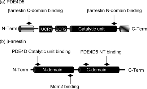
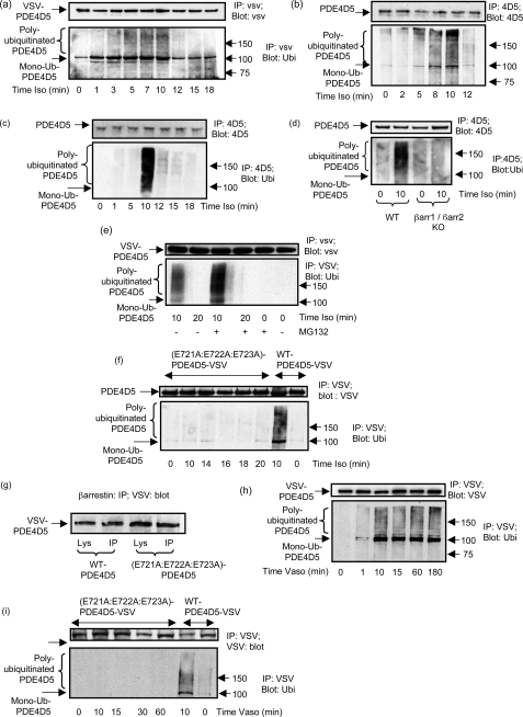
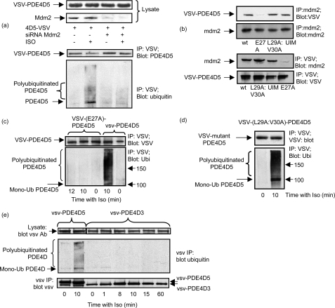
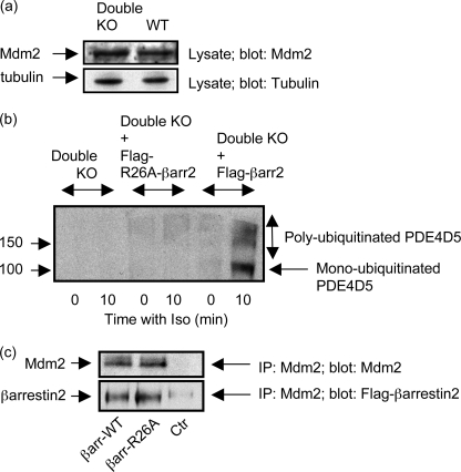
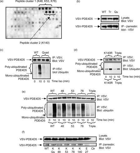
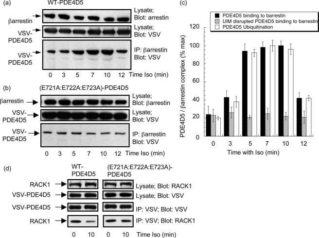
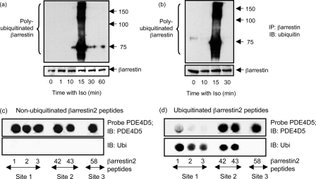
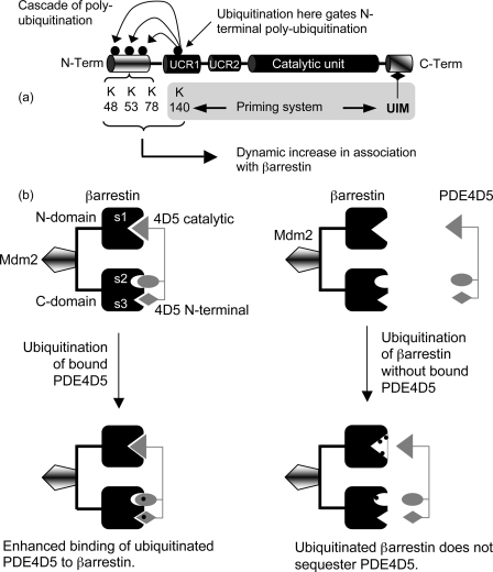
Beta-arrestin plays a key role in regulating beta2-adrenoreceptor signaling by interdicting activation of adenylyl cyclase and selectively sequestering cAMP phosphodiesterase-4D5 (PDE4D5) for delivery of an active cAMP degrading system to the site of cAMP synthesis. Here we show that the beta-agonist, isoprenaline, triggers the rapid and transient ubiquitination of PDE4D5 in primary cardiomyocytes, mouse embryo fibroblasts, and HEK293B2 cells constitutively expressing beta2-adrenoceptors. Reconstitution analyses in beta-arrestin1/2 double knockout cells plus small interference RNA knockdown studies indicate that a beta-arrestin-scaffolded pool of the E3-ubiquitin ligase, Mdm2, mediates PDE4D5 ubiquitination. Critical for this is the ubiquitin-interacting motif located in the extreme C terminus of PDE4D5, which is specific to the PDE4D sub-family. In vitro ubiquitination [corrected] of a PDE4D5 spot-immobilized peptide array, followed by a mutagenesis strategy, showed that PDE4D5 ubiquitination occurs at Lys-48, Lys-53, and Lys-78, which are located within its isoform-specific N-terminal region, as well as at Lys-140 located within its regulatory UCR1 module. We suggest that mono-ubiquitination at Lys-140 primes PDE4D5 for a subsequent cascade of polyubiquitination occurring within its isoform-specific N-terminal region at Lys-48, Lys-53, and Lys-78. PDE4D5 interacts with a non-ubiquitinated beta-arrestin sub-population that is likely to be protected from Mdm2-mediated ubiquitination due to steric hindrance caused by sequestered PDE4D5. Ubiquitination of PDE4D5 elicits an increase in the fraction of PDE4D5 sequestered by beta-arrestin in cells, thereby contributing to the fidelity of PDE4D5-beta-arrestin interaction, as well as decreasing the fraction of PDE4D5 sequestered by the scaffolding protein, RACK1.


Figures

FIGURE 1.
FIGURE 1.
Schematics of PDE4D5 and β-arrestin. a, PDE4D5-specific N-terminal (NT) domain (aa 1–88), the regulatory UCR1 (aa 122–182) and UCR2 (aa 206–284) domains, the catalytic domain (aa 297–683) and the PDE4D sub-family-specific C-terminal (CT) domain (aa 684–745). Indicated are the UIM (aa 721–734; EEEaVgeeeeSqpE), the sites of interaction with β-arrestin as aa 35–88 for the β-arrestin C-domain and aa 670–676 for its N-domain. b, N- (aa 1–180) and C- (aa 188–362) domains of β-arrestin2 with the regions identified for interaction with both PDE4D5 (aa 18–26, aa 215–220, and aa 286–291) and Mdm2 (aa 154–180).
FIGURE 2.
FIGURE 2.
GPCR agonist-stimulated ubiquitination of PDE4D5. a, HEKB2 cells were transfected to express VSV-tagged PDE4D5 and then challenged for the indicated times with isoprenaline (10 μm). VSV-tagged PDE4D5 was immunopurified and immunoblotted with either a VSV antibody or an ubiquitin antibody. b, rat cardiomyocytes were similarly treated, PDE4D5-immunopurified, and then immunoblotted with either an antiserum specific for ubiquitin or one specific for PDE4D5. c, untransfected HEKB2 cells were challenged for the indicated times with isoprenaline (10 μm) and endogenous PDE4D5-immunopurified and then immunoblotted with either an antiserum specific for ubiquitin or one specific for PDE4D5. d, MEFs were treated or not for 10 min with isoprenaline (10 μm) with analysis as in c. e, as in a but, where indicated, cells were also treated with MG132 (50 nm); f, HEKB2 cells were transfected with the UIM-disrupted E721A:E722A:E723A-VSV-PDE4D5 mutant and then challenged for the indicated times with isoprenaline (10 μm) prior to immunopurification of the VSV-tagged PDE4D5 mutant and immunoblotting with either a VSV antibody (upper panel) or an ubiquitin antibody (lower panel). PDE4D5 wild-type also analyzed as a positive control. g, β-arrestin was immunopurified from cells transfected with VSV-tagged versions of either wild-type PDE4D5 of the UIM-disrupted E721A:E722A:E723A-PDE4D5 mutant and both input lysates and immunoprecipitates blotted with antiserum to the VSV epitope tag. h, HEK cells stably overexpressing the vasopressin-V2 receptor were transfected to express VSV-tagged PDE4D5 and then challenged for the indicated times with vasopressin (10 μm) prior to analysis as in a. i, as in h, but here cells were transfected with either wild-type VSV-tagged PDE4D5 or the UIM-disrupted E721A:E722A:E723A-PDE4D5 VSV-tagged mutant and then challenged for the indicated times with vasopressin (10 μm). PDE4D5 wild-type also analyzed as a positive control. Data show experiments typical of ones performed three times.
FIGURE 3.
FIGURE 3.
PDE4D5 ubiquitination requires Mdm2 and β-arrestin. a, HEKB2 cells transfected to express VSV-PDE4D5 either were or were not subjected to small interference RNA-mediated knockdown of Mdm2 (Mdm2 KO) prior to isoprenaline (10 μm) challenge (10 min). Upper panel shows lysate immunoblots for endogenous Mdm2 and VSV-PDE4D5. Lower panel shows immunopurified VSV-PDE4D5 blotted for ubiquitin and VSV. b, HEKB2 cells transfected to express the indicated mutant forms of VSV-PDE4D5 were challenged with isoprenaline and analyzed as in a. HEKB2 cells were transfected to express either wild type or the indicated mutant forms of VSV-tagged PDE4D5. UIM is the UIM-disrupted E721A:E722A:E723A-PDE4D5-VSV mutant; E27A-VSV-PDE4D5 is a mutant that fails to bind β-arrestin; and L29A:V30A-VSV PDE4D5 is a mutant that can bind β-arrestin, but which fails to bind RACK1. Upper panel shows immunopurified endogenous Mdm2 blotted for Mdm2 and for VSV-PDE4D5. Lower panel shows immunopurified VSV-PDE4D5 blotted for Mdm2 and for VSV-PDE4D5. c, HEKB2 cells were transfected to express the E27A-VSV-PDE4D5, a mutant that fails to bind β-arrestin, then challenged for the indicated times with isoprenaline (10 μm) prior to immunopurification of the VSV-tagged PDE4D5 mutant, which was then immunoblotted with either a VSV antibody (upper panel) or an ubiquitin antibody (lower panel). PDE4D5 wild type also analyzed as a positive control. d, as in c but with L29A:V30A-VSV PDE4D5, a mutant that can bind β-arrestin, but which fails to bind RACK1. e, cells were transfected with VSV-tagged forms of either PDE4D5 or PDE4D3 and challenged for the indicated times with isoprenaline prior to analysis as in c. Data show experiments typical of ones performed three times.
FIGURE 4.
FIGURE 4.
β-Arrestin1/2 double knockout mouse embryo fibroblasts. a, equal amounts of lysates (protein) from wild-type MEFs and MEFs from β-arrestin1/2 double knockout animals (Double KO) were blotted for Mdm2. Similar levels were found in both sets of cells as assessed by densitometry (<10% difference). b, double β-arrestin1/2 knockout MEFs (Double KO) either were or were not transfected with FLAG-tagged β-arrestin2 prior to challenge for 10 min with isoprenaline (10 μm). Endogenous PDE4D5 was immunopurified and blotted for PDE4D5 and ubiquitin. In a control “rescue” experiment, double β-arrestin1/2 knockout MEFs (Double KO) either were or were not transfected with the Arg-26 → Ala mutant of FLAG-tagged β-arrestin2, FLAG-tagged (R26A)-β-arrestin2, which does not bind PDE4D5, prior to challenge for 10 min with isoprenaline (10 μm). Endogenous PDE4D5 was then immunopurified and blotted for PDE4D5 and ubiquitin. c, Mdm2 was immunoprecipitated from double knockout MEFS that either had or had not been transfected with FLAG-tagged (R26A)-β-arrestin2, and the immunoprecipitate blotted for either Mdm2 or for FLAG-tagged β-arrestin2. A “beads only” control (Ctr) is also shown where no Mdm2-specific antiserum was used in the immunoprecipitation protocol. Data show experiments typical of ones performed three times.
FIGURE 5.
FIGURE 5.
PDE4D5 ubiquitination sites. a, a scanning peptide array of the entire 745-amino acid sequence of PDE4D5 was generated using overlapping immobilized 25-mer peptides each displaced by 5 residues. This was subjected to in vitro ubiquitination with recombinant Mdm2 and subsequent detection of bound ubiquitin using an anti-ubiquitin antibody. The dark spots indicate positive interacting peptides, whose sequence incorporated the indicated lysine residues. b, HEKB2 cells were transfected with either wild-type VSV-PDE4D5 or the Quad (K48R:K53R:K78R:K140R)-VSV-PDE4D5 or Triple (K48R:K53R:K78R)-VSV-PDE4D5 mutant forms. Lysates expressing equal amounts of each VSV-PDE4D5 species were taken, then β-arrestin immunopurified, and the amount of associated VSV-PDE4D5 species was identified by immunoblotting. Quantitative densitometry showed no difference (<10%) between the amounts of these species sequestered by β-arrestin in resting cells. c–e, HEKB2 cells were transfected to express either wild-type or mutant forms of VSV-tagged PDE4D5 and, where indicated, challenged for 10 min with isoprenaline (10 μm). VSV-PDE4D5 was immunopurified and blotted for VSV and ubiquitin. In c Quad/Qu mutant is (K48R:K53R:K78R:K140R), in d Triple/Tr is (K48R:K53R:K78R) and K140R is Lys-140 → Arg, and in e, 48 is K48R, 53 is K53R, and 78 is K78R. f, HEKB2 cells were transfected with the arginine mutants of indicated residues in VSV-tagged PDE4D5, endogenous β-arrestin was immunoprecipitated using a specific antiserum and the immunoprecipitates blotted with a VSV-specific antiserum to detect PDE4D5. Quad indicates the (K48R:K53R:K78R:K140R)-VSV-PDE4D5 species and Ctr is the beads only control where no β-arrestin was used in the immunoprecipitation (IP) protocol. Data show experiments typical of ones performed three times.
FIGURE 6.
FIGURE 6.
Isoprenaline increases β-arrestin-sequestered PDE4D5. HEKB2 cells were transfected to express either wild-type VSV-PDE4D5 (a) or the UIM-disrupted E721A:E722A:E723A-VSV-PDE4D5 mutant (b) and then challenged with isoprenaline (10 μm) for the indicated times. After this, β-arrestin was immunopurified and blotted for VSV-PDE4D5 with lysates blotted for both β-arrestin and VSV-PDE4D5. c, quantification (mean ± S.D.; n = 3) quantifying isoprenaline-stimulated changes in β-arrestin-sequestered wild-type VSV-PDE4D5 and the UIM-disrupted E721A:E722A:E723A-PDE4D5 mutant and for the level of poly-ubiquitinated wild-type VSV-PDE4D5. d, cells were transfected as in a, and lysates were blotted as indicated for either VSV, to detect recombinant PDE4D5, or for RACK1 and VSV-immunoprecipitates blotted for either PDE4D5-VSV or RACK1. All immunoblotting data show experiments typical of ones performed at least three times.
FIGURE 7.
FIGURE 7.
Ubiquitination of β-arrestin. In a HEKB2 cells were transfected with wild-type-PDE4D5 and then challenged with isoprenaline (10 μm) for the indicated times, whereupon endogenous β-arrestin was selectively immunopurified and gels blotted for ubiquitin (upper panel) or β-arrestin (lower panel). In b cells were transfected with the Quad (K4R) mutant (K48R:K53R:K78R:K140R)-PDE4D5, which does not become ubiquitinated, prior to isoprenaline challenge for the indicated times and subsequent analysis of β-arrestin ubiquitination, as in a. Scanning analyses of β-arrestin peptide arrays have previously identified five peptides that interact with purified recombinant PDE4D5-MBP fusion protein probe expressed in E. coli (38). Five such peptides were synthesized, immobilized, and probed here with recombinant PDE4D5-MBP prior to detection with both an antibody to PDE4D5 (c and d; upper panel) and one to ubiquitin (c and d; lower panel), as indicated, using Odyssey IR-tagged secondary antisera. c, native untreated arrays and d shows arrays subjected to prior in vitro ubiquitination with Mdm2 as the E3 ligase. Data show experiments typical of ones performed three times. Peptide region: 1–3, (1–35) MGEKPGTRVFK(11)K(12)SSPNCKLTVYLGKRDFVDHLDKV; 42 and 43, (206–235) KELYYHGEPLNVNVHVTNNSTKTVKKIKVS; and 58, (286–310) RGLALDGKLKHEDTNLASSTIVKEG. Data show experiments typical of ones performed three times.
FIGURE 8.
FIGURE 8.
Ubiquitination of PDE4D5 and β-arrestin sub-populations. a, a schematic of PDE4D5 with its catalytic unit and isoform-specific N-terminal region, showing the location of its UIM, sites of ubiquitination, and proposing that an initiating/priming ubiquitination occurs at Lys-140 that triggers a cascade of ubiquitination at Lys-48, Lys-53, and Lys-78 within the unique N-terminal region of PDE4D5. b, a schematic proposing that PDE4D5 binds only non-ubiquitinated β-arrestin as ubiquitination within Site 1 on β-arrestin prevents PDE4D5 sequestration. However, isoprenaline challenge allows β-arrestin-sequestered PDE4D5 to be ubiquitinated by sequestered Mdm2, thereby enhancing β-arrestin-PDE4D5 interaction and aiding the fidelity of sequestration of PDE4D5 by β-arrestin.

Similar articles

Cited by

References

    1. Beavo J. A., Brunton L. L. ( 2002) Nat. Rev. Mol. Cell Biol. 3, 710– 718 - PubMed
    1. Taskén K., Aandahl E. M. ( 2004) Physiol. Rev. 84, 137– 167 - PubMed
    1. Taylor S. S., Kim C., Vigil D., Haste N. M., Yang J., Wu J., Anand G. S. ( 2005) Biochim. Biophys. Acta 1754, 25– 37 - PubMed
    1. Wong W., Scott J. D. ( 2004) Nat. Rev. Mol. Cell Biol. 5, 959– 970 - PubMed
    1. Conti M., Beavo J. ( 2007) Annu. Rev. Biochem. 76, 481– 511 - PubMed

Publication types

MeSH terms