. 2009 Jul;23(7):1092-101.
doi: 10.1210/me.2009-0030. Epub 2009 Apr 16.

ERK signaling in the pituitary is required for female but not male fertility

Affiliations

ERK signaling in the pituitary is required for female but not male fertility

Stuart P Bliss et al. Mol Endocrinol. 2009 Jul.

Abstract

Males and females require different patterns of pituitary gonadotropin secretion for fertility. The mechanisms underlying these gender-specific profiles of pituitary hormone production are unknown; however, they are fundamental to understanding the sexually dimorphic control of reproductive function at the molecular level. Several studies suggest that ERK1 and -2 are essential modulators of hypothalamic GnRH-mediated regulation of pituitary gonadotropin production and fertility. To test this hypothesis, we generated mice with a pituitary-specific depletion of ERK1 and 2 and examined a range of physiological parameters including fertility. We find that ERK signaling is required in females for ovulation and fertility, whereas male reproductive function is unaffected by this signaling deficiency. The effects of ERK pathway ablation on LH biosynthesis underlie this gender-specific phenotype, and the molecular mechanism involves a requirement for ERK-dependent up-regulation of the transcription factor Egr1, which is necessary for LHbeta expression. Together, these findings represent a significant advance in elucidating the molecular basis of gender-specific regulation of the hypothalamic-pituitary-gonadal axis and sexually dimorphic control of fertility.


Figures

Figure 1
Figure 1
Validation of the ERK1/2 DKO mouse. A, Cre-mediated recombination at the ERK2 locus within the pituitary was detected by PCR using genomic DNA from the specified tissues. For each DNA sample, the forward primer designated “F” was paired individually with reverse primers labeled “5,” “7,” and “9” spanning the floxed genomic region as indicated in the schematic. Lanes are labeled with the specified reverse primer used for the reaction. Molecular weight marker is shown in the left lane. Equivalent results were obtained from males and females. B, Cell type-specific loss of ERK protein in the ERK1/2 DKO was determined by coimmunofluorescent labeling of pituitary sections from control (panels A–C) and DKO (panels D–F) male animals using fluorescein isothiocyanate-conjugated antibodies against ERK1/2 and Texas Red-conjugated antibodies against LH-β. ERK (A and D), LHβ (B and E), and simultaneous visualization of both wavelengths (C and F) are shown. Arrows indicate representative LHβ-positive cells with distinct lack of ERK labeling. Bars, 100 μm.
Figure 2
Figure 2
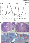
Assessment of estrous cycle activity in the ERK1/2 DKO mouse. A, Vaginal cytology was performed at 24-h intervals by microscopic examination of Wright’s stained vaginal lavage effluents. One hundred cells were counted, and the percentage of epithelial cells at each time point was determined. Data are shown for a single animal; however, similar results were obtained from three animals of each genotype. B, Hematoxylin and eosin-stained histological sections of ovaries from control (panel A) and DKO (panel B) animals are shown. A representative corpus luteum is indicated (CL). Microscopic sections from control (panel C), and DKO (panel D) ovaries were immunohistochemically stained using an antibody against ERK1/2. Both control and DKO animals were ERK1 null; thus all specific labeling represents ERK2 protein. A and B, bars, 200 μm; C and D, bars, 100 μm.
Figure 3
Figure 3
Evaluation of thyroid function in the ERK1/2 DKO mouse. A, Relative transcript levels of TSHβ from whole pituitaries of female control and DKO animals were determined by quantitative PCR. Results were calibrated to levels of TSHβ mRNA from pituitaries of randomly cycling female wild-type mice. Bars represent mean ± sem for five animals of each genotype. Means were compared by two-tailed t test. B, Levels of TSHβ in whole pituitary lysates from control and DKO animals were compared by immunoblot. Actin is shown as a lane-loading control. C, Serum levels of total T4 from randomly cycling wild-type, control, and DKO females were determined by ELISA. Bars represent mean ± sem for three (wild type) or 11 (control and DKO) animals in the respective groups. Means for control and DKO groups were compared by two-tailed t test.
Figure 4
Figure 4
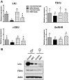
Basal expression of gonadotropin subunit and GnRHR genes in the ERK1/2 DKO mouse. A, Whole pituitary relative transcript levels of the specified genes were determined by quantitative PCR. Results were calibrated to corresponding transcript levels in pituitaries of randomly cycling female wild-type mice. Bars represent mean ± sem for five animals of each gender and genotype. Bars not sharing common letter designations represent mean values that are statistically significantly different (P < 0.05). B, Levels of LHβ and FSHβ protein in whole pituitary lysates from control and DKO animals were compared by immunoblotting. Actin is shown as a lane-loading control.
Figure 5
Figure 5
Pharmacological superovulation rescues the anovulatory phenotype of the ERK1/2 DKO mouse. Control and DKO females were injected ip with vehicle or 5 U pregnant mare serum gonadotropin, followed in 48 h by vehicle or 5 U of human chorionic gonadotropin. After 9 d, ovaries were collected and examined histologically. Ovarian sections from vehicle-treated (A and B) and superovulated (C and D) control (A and C) and DKO (B and D) animals are shown. Arrows indicate corpora lutea. Bars, 200 μm.
Figure 6
Figure 6
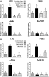
ERK1/2 DKO animals of both genders fail to up-regulate LHβ after gonadectomy. Control and DKO animals were either castrated (Csx), ovariectomized (Ovx), or sham operated (sham). After 7 d, pituitaries were collected and analyzed by qPCR for relative transcript levels of the indicated gonadotropin subunit genes and the GnRHR. Data are shown separately for males (A) and females (B). Results were calibrated to corresponding transcript levels in pituitaries of randomly cycling female wild-type mice. Bars represent mean ± sem for six males or seven females per group and represent pooled results from two separate experiments. Bars not sharing common letter designations represent mean values that are statistically significantly different (P < 0.05). Note the differences in scaling of the y-axis for the FSHβ results in the female.
Figure 7
Figure 7
Serum gonadotropin levels do not increase in DKO animals of either gender after gonadectomy. Control and DKO animals were either castrated (Csx), ovariectomized (Ovx), or sham operated (sham). After 7 d, animals were euthanized, and serum levels of LHβ and FSHβ were determined by ELISA. Data are shown separately for males (A) and females (B). Bars represent mean ± sem for six males or seven females per group and represent pooled results from two separate experiments. Bars not sharing common letter designations represent mean values that are statistically significantly different (P < 0.05).
Figure 8
Figure 8
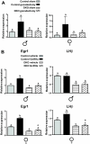
ERK-deficient gonadotropes fail to up-regulate the immediate early gene Egr1 in response to GnRH stimulation. A, Control and DKO animals of both genders were either gonadectomized (gonadectomy) or sham operated (sham). After 7 d, pituitaries were collected and analyzed by qPCR for relative transcript levels of Egr1. Data are shown separately for males (left panel) and females (right panel). Results were calibrated to corresponding transcript levels in pituitaries of randomly cycling female wild-type mice. Bars represent mean ± sem for six animals per group and represent pooled results from two separate experiments for each gender. Bars not sharing common letter designations represent mean values that are statistically significantly different (P < 0.05). Pituitaries from control and DKO animals were dispersed into primary culture (two pituitaries per well) and treated for 20 min with vehicle or with 100 nm of the GnRH agonist buserelin (GnRHa). Relative transcript levels of Egr1 were measured in each sample by qPCR. Bars represent mean ± sem for six wells representing pooled results from two separate experiments. Data are shown separately for males (left) and females (right). For these experiments, the expression level of the vehicle-treated control animals was arbitrary assigned as the calibrator.

References

    1. Pearson G, Robinson F, Beers Gibson T, Xu BE, Karandikar M, Berman K, Cobb MH 2001 Mitogen-activated protein (MAP) kinase pathways: regulation and physiological functions. Endocr Rev 22:153–183 - PubMed
    1. Mazzucchelli C, Vantaggiato C, Ciamei A, Fasano S, Pakhotin P, Krezel W, Welzl H, Wolfer DP, Pagès G, Valverde O, Marowsky A, Porrazzo A, Orban PC, Maldonado R, Ehrengruber MU, Cestari V, Lipp HP, Chapman PF, Pouysségur J, Brambilla R 2002 Knockout of ERK1 MAP kinase enhances synaptic plasticity in the striatum and facilitates striatal-mediated learning and memory. Neuron 34:807–820 - PubMed
    1. Nekrasova T, Shive C, Gao Y, Kawamura K, Guardia R, Landreth G, Forsthuber TG 2005 ERK1-deficient mice show normal T cell effector function and are highly susceptible to experimental autoimmune encephalomyelitis. J Immunol 175:2374–2380 - PubMed
    1. Yao Y, Li W, Wu J, Germann UA, Su MS, Kuida K, Boucher DM 2003 Extracellular signal-regulated kinase 2 is necessary for mesoderm differentiation. Proc Natl Acad Sci USA 100:12759–12764 - PMC - PubMed
    1. Newbern J, Zhong J, Wickramasinghe SR, Li X, Wu Y, Samuels I, Cherosky N, Karlo JC, O'Loughlin B, Wikenheiser J, Gargesha M, Doughman YQ, Charron J, Ginty DD, Watanabe M, Saitta SC, Snider WD, Landreth GE 2008 Mouse and human phenotypes indicate a critical conserved role for ERK2 signaling in neural crest development. Proc Natl Acad Sci 105:17115–17120 - PMC - PubMed

Publication types

MeSH terms

Substances