. 2009 Jun;37(11):3723-38.
doi: 10.1093/nar/gkp229. Epub 2009 Apr 16.

Molecular mechanism of poly(ADP-ribosyl)ation by PARP1 and identification of lysine residues as ADP-ribose acceptor sites

Affiliations

Molecular mechanism of poly(ADP-ribosyl)ation by PARP1 and identification of lysine residues as ADP-ribose acceptor sites

Matthias Altmeyer et al. Nucleic Acids Res. 2009 Jun.

Abstract

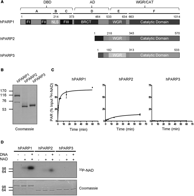
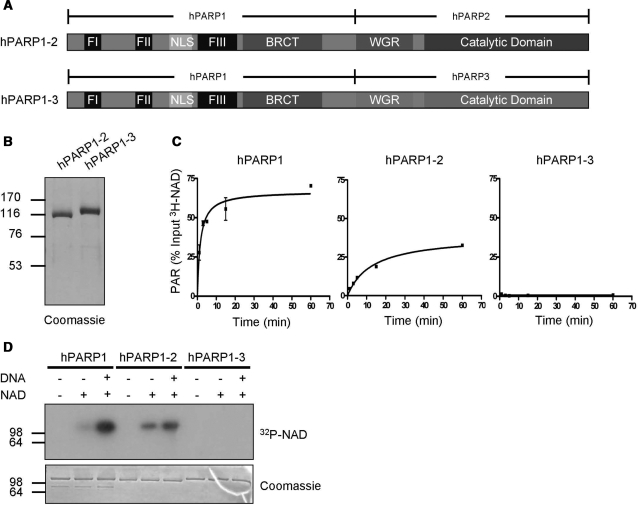
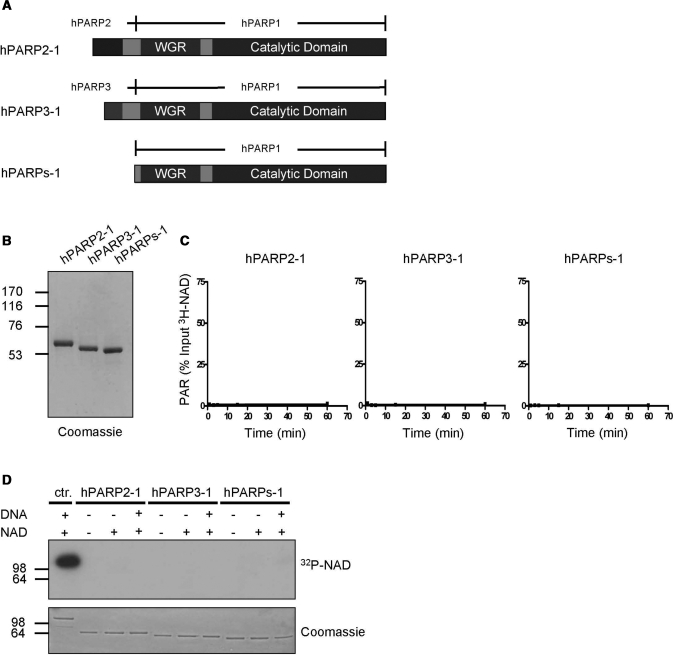
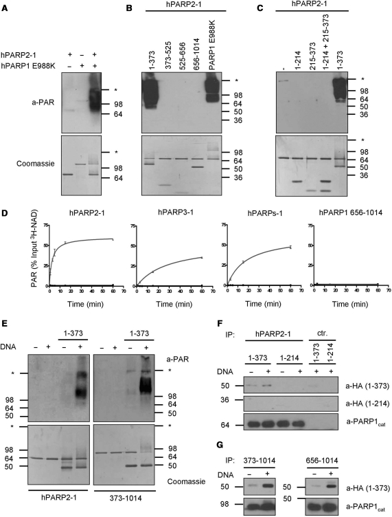
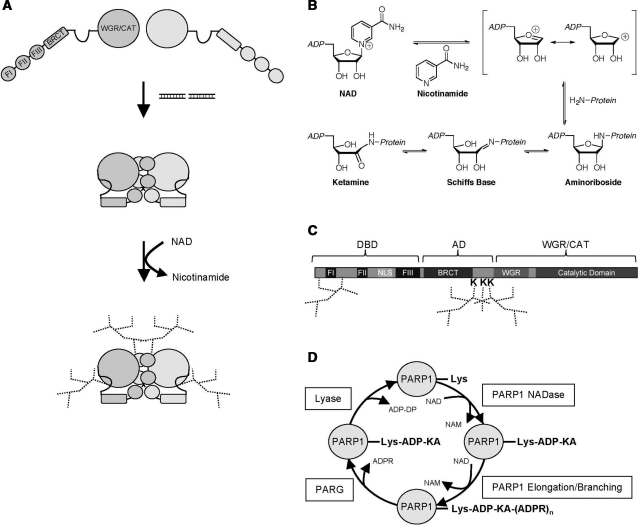
Poly(ADP-ribose) polymerase 1 (PARP1) synthesizes poly(ADP-ribose) (PAR) using nicotinamide adenine dinucleotide (NAD) as a substrate. Despite intensive research on the cellular functions of PARP1, the molecular mechanism of PAR formation has not been comprehensively understood. In this study, we elucidate the molecular mechanisms of poly(ADP-ribosyl)ation and identify PAR acceptor sites. Generation of different chimera proteins revealed that the amino-terminal domains of PARP1, PARP2 and PARP3 cooperate tightly with their corresponding catalytic domains. The DNA-dependent interaction between the amino-terminal DNA-binding domain and the catalytic domain of PARP1 increased V(max) and decreased the K(m) for NAD. Furthermore, we show that glutamic acid residues in the auto-modification domain of PARP1 are not required for PAR formation. Instead, we identify individual lysine residues as acceptor sites for ADP-ribosylation. Together, our findings provide novel mechanistic insights into PAR synthesis with significant relevance for the different biological functions of PARP family members.


Figures

Figure 1.
Figure 1.
Purified full-length human PARP1 and PARP2 are enzymatically active. (A) Domain organization of human PARP1, PARP2 and PARP3. Letters A–F indicate domain nomenclature of PARP1 and numbers indicate amino acid positions. (B) Purity of PARP family members after one step affinity chromatography. One microgram of each recombinant, purified protein was used for SDS–PAGE followed by coomassie staining. (C) Time course of PAR formation by different PARP family members. 3H-NAD incorporation into TCA-precipitable polymers was determined by scintillation counts. Substrate concentration: 400 μM 3H-NAD. Reactions were performed in triplicates, error bars represent standard deviations. (D) Auto-modification of different PARP family members detected by autoradiography. Substrate concentration: 100 nM 32P-NAD. Molecular size markers in kilo Daltons are indicated.
Figure 2.
Figure 2.
The carboxyl-terminal domains of PARP1, PARP2 and PARP3 cannot compensate for each other. (A) Domain organization of chimera PARP1-2 and chimera PARP1-3. (B) Purity of chimera PARP1-2 and chimera PARP1-3 after one-step affinity chromatography. One microgram of each recombinant, purified protein was used for SDS–PAGE followed by coomassie staining. (C) Time course of PAR formation by PARP1, chimera PARP1-2 and chimera PARP1-3 as in Figure 1C. (D) Auto-modification of PARP1, chimera PARP1-2 and chimera PARP1-3 detected by autoradiography as in Figure 1D. Molecular size markers in kilo Daltons are indicated.
Figure 3.
Figure 3.
The carboxyl-terminal domain of PARP1 is not activated by the amino-terminal domains of PARP2 or PARP3. (A) Domain organization of chimera PARP2-1, chimera PARP3-1 and PARPs-1. (B) Purity of chimera PARP2-1, chimera PARP3-1 and PARPs-1 after one-step affinity chromatography. One microgram of each recombinant, purified protein was used for SDS–PAGE followed by coomassie staining. (C) Time course of PAR formation by chimera PARP2-1, chimera PARP3-1 and PARPs-1 as in Figure 1C. (D) Auto-modification of PARP1 (ctr.), chimera PARP2-1, chimera PARP3-1 and PARPs-1 detected by autoradiography as in Figure 1D. Molecular size markers in kilo Daltons are indicated.
Figure 4.
Figure 4.
The DBD of PARP1 interacts with and is sufficient to stimulate its WGR/CAT domain. (A) PAR formation by chimera PARP2-1 co-incubated with catalytically inactive PARP1 E988K. PAR was detected by western blot using anti-PAR antibody LP96-10. Substrate concentration: 400 μM NAD. (B) PAR formation by chimera PARP2-1 co-incubated with the indicated fragments of PARP1 or with PARP1 E988K. (C) PAR formation of chimera PARP2-1 co-incubated with the indicated fragments or combination of fragments of PARP1. (D) Time course of PAR formation by chimera PARP2-1, chimera PARP3-1, PARPs-1 and PARP1 656–1014 in the absence or presence of fragment 1–373 as in Figure 1C. Black without fragment 1–373 and grey with fragment 1–373. (E) PAR formation of chimera PARP2-1 or PARP1 373–1014 co-incubated with fragment 1–373 in the absence or presence of DNA. (F) In vitro interaction between chimera PARP2-1 and 1–373. Chimera PARP2-1 was bound to protein A sepharose using an antibody against the CAT of PARP1 (a-PARP1cat) and was then incubated with HA-tagged fragment 1–373 or 1–214 in the absence or presence of DNA. HA-tagged fragments were detected by western blot. PARP1cat antibody coupled to beads without chimera PARP2-1 served as control (ctr.). (G) In vitro interaction between PARP1 373–1014 or 656–1014 with 1–373. Experiments were performed as described in (F). Molecular size markers in kilo Daltons and the border between stacking and separating gel (asterisk) are indicated.
Figure 5.
Figure 5.
PARP1 forms a catalytic dimer which requires at least one functional FI and FIII domain for activity. (A) Domain organization of the PARP1 deletion mutants used for this figure. (B) PAR formation by PARP1 when co-incubated with the indicated inactive proteins or fragments at a molar ratio of 1:1 or 1:5. According to the manufacturer, the anti-PAR antibody LP96-10 cross reacts with bovine serum albumin (BSA) (band at around 64 kDa). (C) PAR formation by DBD deletion mutants ΔFI, ΔFII and ΔFIII. (D) PAR formation by a combination of the two DBD deletion mutants ΔFI and ΔFIII. (E) PAR formation by the DBD deletion mutants ΔFI and ΔFIII when they were co-incubated with catalytically inactive PARP1 mutants or with fragment 1–373. (F) PAR formation by PARP1 lacking the WGR domain. (G) PAR formation by PARP1 ΔWGR in combination with DBD deletion mutants, catalytically inactive PARP1 mutants or with fragment 1–373. Molecular size markers in kilo Daltons and the border between stacking and separating gel (asterisk) are indicated.
Figure 6.
Figure 6.
Lysine residues within the AD of PARP1 are target sites for poly(ADP-ribosyl)ation. (A) Domain organization of the PARP1 mutants used for this figure. (B) Auto-modification of the indicated PARP1 mutants lacking either the BRCT domain (ΔBRCT) or the BRCT domain and carrying substitutions for all glutamic acid residues in the remaining stretch of the AD (ΔBRCT/E). (C) Auto-modification of a PARP1 deletion mutant lacking aa 466–525 (ΔAc), a region that was previously shown to be acetylated. (D) Trans-poly(ADP-ribosyl)ation of the AD from amino acid 373–525 by PARP1. KTR, K498/521/524R. (E) Auto-modification of a PARP1 K498/521/524R mutant. Molecular size markers in kilo Daltons are indicated.
Figure 7.
Figure 7.
Model for PARP1 activation and ADP-ribosylation of lysine residues. (A) Model for the sequential activation and regulation of PARP1. The DNA-dependent interaction between the DBD and the WGR/CAT induces a state of high substrate affinity and high turnover rate in PARP1. Subsequently, acceptor amino acids in the auto-modification loop as well as in the DBD are poly(ADP-ribosyl)ated. (B) Proposed reaction mechanism for NADase-dependent auto-ADP-ribosylation of lysine residues by PARP1 via Schiff base formation. (C) Scheme depicting the identified PAR acceptor lysine residues in PARP1. (D) A revised view of ADP-ribose metabolism. PARP1 catalyzes lysine mono(ADP-ribosyl)ation via its NADase activity and subsequently PAR chain elongation. PARG cleaves glycosidic ribose–ribose bonds to generate PARP1-Lys-ADP-KA, which is then substrate for an ADP-ribosyl protein lyase. See discussion for details. NAM, nicotinamide; Lys, lysine; ADP-KA, ADP-ketamine; ADPR, ADP-ribose; ADP-DP, ADP-3″-deoxypentose-2″-ulose.

Similar articles

Cited by

References

    1. Hassa PO, Hottiger MO. The diverse biological roles of mammalian PARPS, a small but powerful family of poly-ADP-ribose polymerases. Front. Biosci. 2008;13:3046–3082. - PubMed
    1. Hassa PO, Haenni SS, Elser M, Hottiger MO. Nuclear ADP-ribosylation reactions in mammalian cells: where are we today and where are we going? Microbiol. Mol. Biol. Rev. 2006;70:789–829. - PMC - PubMed
    1. Suzuki H, Uchida K, Shima H, Sato T, Okamoto T, Kimura T, Miwa M. Molecular cloning of cDNA for human poly(ADP-ribose) polymerase and expression of its gene during HL-60 cell differentiation. Biochem. Biophys. Res. Commun. 1987;146:403–409. - PubMed
    1. Cherney BW, McBride OW, Chen DF, Alkhatib H, Bhatia K, Hensley P, Smulson ME. cDNA sequence, protein structure, and chromosomal location of the human gene for poly(ADP-ribose) polymerase. Proc. Natl Acad. Sci. USA. 1987;84:8370–8374. - PMC - PubMed
    1. Kameshita I, Matsuda M, Nishikimi M, Ushiro H, Shizuta Y. Reconstitution and poly(ADP-ribosyl)ation of proteolytically fragmented poly(ADP-ribose) synthetase. J. Biol. Chem. 1986;261:3863–3868. - PubMed

Publication types

MeSH terms