. 2009 Jun;37(11):3699-713.
doi: 10.1093/nar/gkp233. Epub 2009 Apr 16.

Histone deacetylase Rpd3 antagonizes Sir2-dependent silent chromatin propagation

Affiliations

Histone deacetylase Rpd3 antagonizes Sir2-dependent silent chromatin propagation

Jing Zhou et al. Nucleic Acids Res. 2009 Jun.

Abstract

In the eukaryotic genome, transcriptionally silent chromatin tends to propagate along a chromosome and encroach upon adjacent active chromatin. The silencing machinery can be stopped by chromatin boundary elements. We performed a screen in Saccharomyces cerevisiae for proteins that may contribute to the establishment of a chromatin boundary. We found that disruption of histone deacetylase Rpd3p results in defective boundary activity, leading to a Sir-dependent local propagation of transcriptional repression. In rpd3 Delta cells, the amount of Sir2p that was normally found in the nucleolus decreased and the amount of Sir2p found at telomeres and at HM and its adjacent loci increased, leading to an extension of silent chromatin in those areas. In addition, Rpd3p interacted directly with chromatin at boundary regions to deacetylate histone H4 at lysine 5 and at lysine 12. Either the mutation of histone H4 at lysine 5 or a decrease in the histone acetyltransferase (HAT) activity of Esa1p abrogated the silencing phenotype associated with rpd3 mutation, suggesting a novel role for the H4 amino terminus in Rpd3p-mediated heterochromatin boundary regulation. Together, these data provide insight into the molecular mechanisms for the anti-silencing functions of Rpd3p during the formation of heterochromatin boundaries.


Figures

Figure 1.
Figure 1.
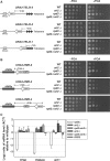
Rpd3L complex is required for establishment of heterochromatic boundaries. (A) Schematic diagrams of the chromosomal locations of three boundary-proximal genes, YPS6 and YIR042C, which are adjacent to telomere, and GIT1, which is proximal to HMR. The HMR-tRNAThr gene and STAR (sub-telomeric anti-silencing region) sequence are also labeled. (B) qRT–PCR results of mRNA levels of boundary-proximal genes, YPS6, YIR042C and GIT1, in wild type and various mutant strains. (C) qRT–PCR results of mRNA levels of YPS6, YIR042C and GIT1 in the individual component deletion mutants of Rpd3L and Rpd3S. Fold transcription is relative to wild type. The log2 ratio less than zero indicates repression of transcription, whereas greater than zero indicates enhancement of transcription. Error bars represent standard error of the mean for three independent RNA purifications. (D) Mating assay to test the effects of mutation of RPD3 on boundary activity of HMR-tRNA. The ∼1.0 kb region flanking the right side of HMR with the HMR tRNAThr boundary gene was cloned into the a2 gene, and these constructs (pRO363 or pRO466) were integrated into chromosome III in a MATα strain in the presence or absence of Rpd3p (see ‘Materials and Methods’ section). The resulting strains and a MATa strain were mated, serially diluted and spotted onto a YC plate or a Ura–/Lys– plate, followed by incubation at 30°C. The photograph was taken after 48 h.
Figure 2.
Figure 2.
Enhanced silencing associated with RPD3 deletion is Sir2-dependent. (A) and (B) The wild-type, rpd3Δ, sir2Δ and rpd3Δ sir2Δ cells harboring a URA3 gene integrated at the subtelomeric regions of chromosome IX (TELIX) or HMR-adjacent loci were serially diluted and spotted onto YC medium with or without 5′-FOA, followed by incubation at 30°C. Photographs were taken after 48 or 72 h. The locations of the integrated URA3 gene on the chromosomes are illustrated on left. The numbers designated as 1–6 represent different positions of the URA3 marker. (C) qRT–PCR results of mRNA levels of YPS6, YIR042C and GIT1 genes in rpd3Δ, sir2Δ, sir3Δ, rpd3Δ sir2Δ, rpd3Δ sir3Δ and RPD3 cells. Fold increase in mRNA is relative to wild type. Error bars represent standard error of the mean for three trials.
Figure 3.
Figure 3.
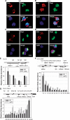
Inactivation of Rpd3p causes redistribution of Sir2p. (A) and (B) Confocal images of the immunolocalization of Sir2p and Rap1p in wild type (A) and rpd3Δ (B) cells. A Sir2p–13Myc fusion protein was stained by mouse anti-Myc monoclonal antibody, detected by a Cy3-conjugated secondary antibody. Rap1p was stained by rabbit anti-Rap1 polyclonal antibody, and detected by a FITC-conjugated secondary antibody. Overlap of these two signals is yellow. DNA is stained by DAPI. The bar indicates 2.0 µm. (C) and (D) Immunolocalization of Sir2p–13Myc and Nop1p in wild-type (C) and rpd3Δ (D) cells. Sir2p–13Myc was stained by rabbit anti-Myc antibody, detected by a Cy3-conjugated secondary antibody. Nop1p was stained by mouse anti-Nop1 antibody, and detected by a FITC-conjugated secondary antibody. DNA is stained by DAPI. (E), (F) and (G) 13Myc–Sir2p binding to rDNA (E), HMR (F) and subtelomeric (G) loci was assayed by ChIP using anti-Myc antibody in wild type and rpd3Δ cells. Average relative Sir2p enrichments are shown for each primer set with its amplified region as indicated on the upper panel of each graph. The upper diagrams in (E), (F) and (G) show a representative portion of ribosomal DNA locus on chromosome XII, two prominent boundary regions in the HMR silent mating type cassette and subtelomeric loci found on chromosome IX-R, respectively (41). Two well-characterized boundary elements, the HMR-adjacent tRNAThr gene and STAR sequence of Chr-IX-R are also shown (8,12,14). Locations of primer sets used for ChIP analysis are designated by the distance to the start coden of HMRA1 gene, or to the start of telomeric X element (∼700 bp to telomere TG1-3 repeats sequence) of chromosome IX. The qPCR data were normalized to an internal control (ARO1) and the input DNA. The results are average of three independent ChIPs with error bars shown for the standard error of the mean for three independent experiments.
Figure 4.
Figure 4.
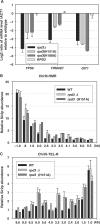
Enzymatic activity of Rpd3p is required for its anti-silencing function. (A) qRT–PCR results of transcription level of boundary-proximal genes, YPS6, YIR042C and GIT1, in wild type and rpd3 mutant cells. Fold transcription relative to wild type are plotted on logarithmic scales. (B) and (C) ChIP assay to detect the Sir2p enrichments at HMR and subtelomeric regions of chromosome IX-R in wild-type and rpd3 mutant strains. Average relative Sir2p enrichments are shown for each primer set with its amplified region denoted as in Figure 3. The data were normalized to an internal control (ARO1) and the input DNA. Error bars represent the standard error of the mean for three independent experiments.
Figure 5.
Figure 5.
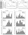
Rpd3p interacts with chromatin at boundary regions to deacetylate histone H4K5 and K12. (A) Protein level of histone Ac-H3, Ac-H4, Ac-H4K5, K8, K12 and K16 in wild-type and rpd3Δ cells was determined by immunoblotting using anti-Ac-H3, anti-Ac-H4, anti-Ac-H4K5, anti-Ac-H4K8, anti-Ac-H4K12 and anti-H4 antibodies, respectively. (B) and (C) Binding of Rpd3p to HMR (B) and subtelomeric regions of chromosome IX (C) was detected by ChIP assay. The qPCR data is normalized to a region approximately 500 bp from the end of chromosome VI-R (TEL 0.5), whereas Rpd3 binding is excluded (45). (D) and (E) Deletion of RPD3 resulted in enhanced acetylation on H4K5 and K12 at HMR-proximal (D) or subtelomeric chromatin (E). For ChIP assay, antibodies against acetylated lysines (Ac-K5, Ac-K8 and Ac-K12) of the H4 histone tail were used. The qPCR data were normalized to an internal control (TEL 0.5) and the input DNA. Average relative enrichments of Ac-H4K5, Ac-H4K8, Ac-H4K12, 13Myc-Rpd3 and no-tag control are shown for each primer set with its amplified region denoted as in Figure 3. The results are average of three independent ChIPs with error bars representing the standard error of the mean for three independent experiments.
Figure 6.
Figure 6.
Deacetylation of H4K5 by Rpd3p is required for antagonizing heterochromatic silencing. (A) The growth phenotype of mutation of histone H4 lysine residues combined with rpd3Δ on subtelomeric URA3 silencing was examined. 10-fold serial dilutions of each yeast cell were spotted on YC plates with 5’-FOA as indicated. (B) qRT–PCR results of transcription level of boundary proximal genes YPS6, YIR042C, and GIT1 genes in wild-type, H4 K5Q mutant, and H4 K5Q and rpd3Δ double mutant cells. Fold transcription is relative to wild-type and plotted on logarithmic scales. (C) and (D) 13Myc–Sir2p binding was assayed by ChIP using anti-Myc antibody in wild-type, H4K5 mutant and H4K5 and rpd3 double mutant cells, at HMR-proximal loci (C) and subtelomeric regions of chromosome IX-R (D). The qPCR data were normalized to an internal control (ARO1) and the input DNA. Average relative Sir2p enrichments are shown for each primer set with its amplified region denoted as in Figure 3. The results presented are an average of three independent ChIPs with error bars shown for standard error. (E) Acetylation of histone H4K5 was measured by immunoblotting in wild-type and eas1(L327S) mutant cells. Histone H4 was used as internal control. (F) The URA3 silencing assay was performed on ESA1, ESA1 rpd3Δ and eas1(L327S) rpd3Δ double mutant cells. Tenfold serial dilutions of each yeast cells were spotted on YC plate with 5′-FOA as indicated.
Figure 7.
Figure 7.
Inactivation of Rpd3p enhances the deposition of H2A.Z at HMR boundary loci. ChIP assay was performed to determine if the 3HA tagged H2A.Z was enriched at boundary loci in wild-type and rpd3Δ cells. A PCR product corresponding to the middle of the open reading frame of PRP8, a gene for which is suggested to be excluded for H2A.Z binding, is used as the internal control. The chromosomal locations of primers were as indicated in the upper panel. The immunoprecipitation data were normalized to input DNA. The results are average of three independent experiments with error bars shown for standard error.

Similar articles

Cited by

References

    1. Richards EJ, Elgin SC. Epigenetic codes for heterochromatin formation and silencing: rounding up the usual suspects. Cell. 2002;108:489–500. - PubMed
    1. Moazed D. Common themes in mechanisms of gene silencing. Mol. Cell. 2001;8:489–498. - PubMed
    1. Nimmo ER, Cranston G, Allshire RC. Telomere-associated chromosome breakage in fission yeast results in variegated expression of adjacent genes. EMBO J. 1994;13:3801–3811. - PMC - PubMed
    1. Allshire RC, Javerzat JP, Redhead NJ, Cranston G. Position effect variegation at fission yeast centromeres. Cell. 1994;76:157–169. - PubMed
    1. Vermaak D, Ahmad K, Henikoff S. Maintenance of chromatin states: an open-and-shut case. Curr. Opin. Cell Biol. 2003;15:266–274. - PubMed

Publication types

MeSH terms