

Deferoxamine reduces intracerebral hematoma-induced iron accumulation and neuronal death in piglets
- PMID: 19372448
- PMCID: PMC2693321
- DOI: 10.1161/STROKEAHA.108.539536
Deferoxamine reduces intracerebral hematoma-induced iron accumulation and neuronal death in piglets
Abstract
Background and purpose: Our previous studies found that deferoxamine reduces intracerebral hemorrhage (ICH)-induced brain injury in rats. The current study examined whether deferoxamine reduces brain injury in a piglet ICH model.
Methods: Pigs received an injection of autologous blood into the right frontal lobe. Deferoxamine (50 mg/kg, IM) or vehicle was administered 2 hours after ICH and then every 12 hours up to 7 days. Animals were killed 3 or 7 days later to examine iron accumulation, white matter injury, and neuronal death.
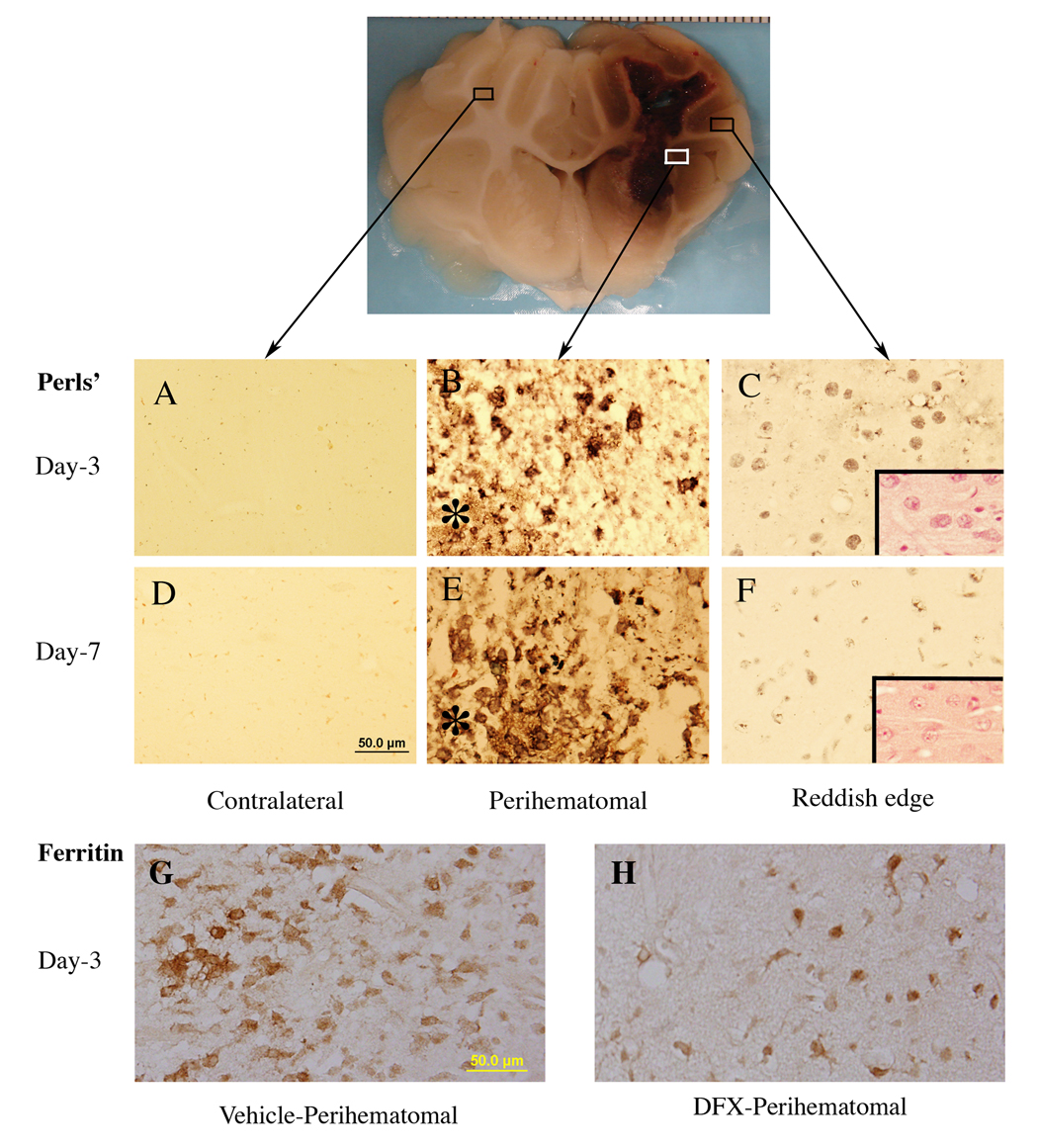
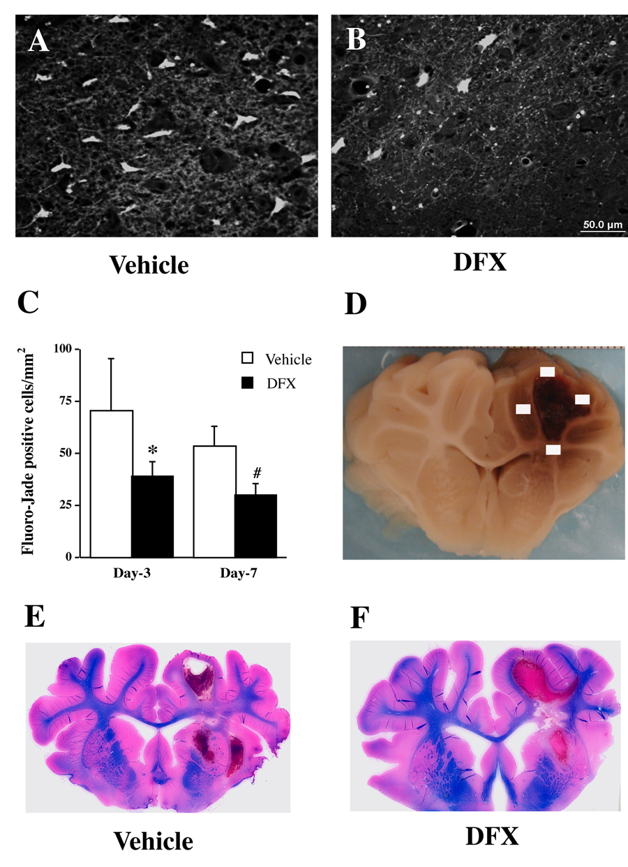
Results: ICH resulted in development of a reddish perihematomal zone, and iron accumulation, ferritin upregulation, and neuronal death within that zone. Deferoxamine reduced the perihematomal reddish zone, white matter injury, and the number of Perls', ferritin, and Fluoro-Jade C-positive cells.
Conclusions: Iron accumulation occurs in the piglet brain after ICH. Deferoxamine reduces ICH-induced iron buildup and brain injury in piglets.
Figures

References
-
- Wagner KR, Sharp FR, Ardizzone TD, Lu A, Clark JF. Heme and iron metabolism: Role in cerebral hemorrhage. J Cereb Blood Flow Metab. 2003;23:629–652. - PubMed
-
- Xi G, Keep RF, Hoff JT. Mechanisms of brain injury after intracerebral hemorrhage. Lancet Neurol. 2006;5:53–63. - PubMed
-
- Nakamura T, Keep RF, Hua Y, Schallert T, Hoff JT, Xi G. Deferoxamine-induced attenuation of brain edema and neurological deficits in a rat model of intracerebral hemorrhage. J Neurosurg. 2004;100:672–678. - PubMed
-
- Hua Y, Nakamura T, Keep RF, Wu J, Schallert T, Hoff JT, Xi G. Long-term effects of experimental intracerebral hemorrhage: The role of iron. J Neurosurg. 2006;104:305–312. - PubMed
-
- Song S, Hua Y, Keep RF, Hoff JT, Xi G. A new hippocampal model for examining intracerebral hemorrhage-related neuronal death: Effects of deferoxamine on hemoglobin-induced neuronal death. Stroke. 2007;38:2861–2863. - PubMed
Publication types
MeSH terms
Substances
Grants and funding
LinkOut - more resources
Full Text Sources
Medical
