

Titration of variant HSV1-tk gene expression to determine the sensitivity of 18F-FHBG PET imaging in a prostate tumor
- PMID: 19372484
- PMCID: PMC2805178
- DOI: 10.2967/jnumed.108.058438
Titration of variant HSV1-tk gene expression to determine the sensitivity of 18F-FHBG PET imaging in a prostate tumor
Abstract
Because of its high selectivity and specificity for the imaging reporter probe 9-(4-(18)F-fluoro-3-[hydroxymethyl]butyl)guanine ((18)F-FHBG), the herpes simplex virus type 1 thymidine kinase (HSV1-tk) variant sr39tk is actively being studied as a PET reporter gene. We recently demonstrated the capability of using a prostate-specific transcriptional amplification PET reporter vector, AdTSTA-sr39tk, to target prostate cancer lymph node metastasis. However, one area that warrants further study is the examination of the sensitivity of PET by determining the minimum percentage of cells expressing the sr39tk transgene needed for detection. Addressing this question could determine the sensitivity of vector-mediated sr39tk PET in cancer-targeting strategies.
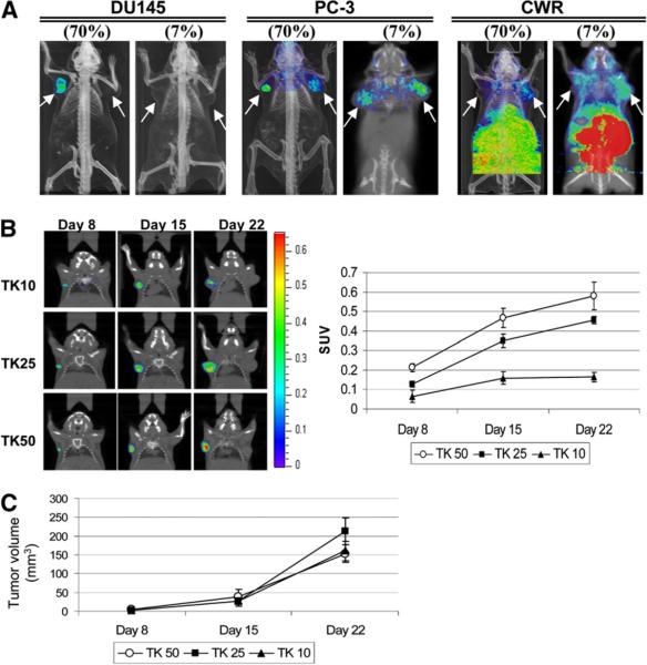
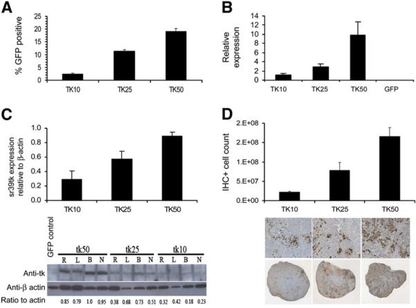
Methods: DU-145, PC-3, and CWR22Rv.1 prostate cancer cell lines (a total of 1 x 10(6) cells) were studied, of which 7%, 10%, 25%, 50%, or 70% were transduced with the lentiviral vector constitutively expressing HSV1-sr39tk-IRES-enhanced green fluorescent protein (EGFP). Cells were subcutaneously implanted into the left shoulder of severe combined immunodeficient mice and evaluated. Tumor cells comparably transduced with an EGFP control vector were implanted on the right shoulder. Mice were imaged using PET with (18)F-FHBG at 8, 15, and 22 d after tumor implant. On day 23, tumors were isolated and analyzed for sr39tk transgene expression by quantitative reverse-transcriptase polymerase chain reaction (RT-PCR), Western blotting, immunohistochemistry, and flow cytometry for EGFP expression.
Results: Results showed a linear relationship between the level of sr39tk expression and the quantity of tracer accrual in DU-145, with the minimal value for PET detection at 10%. The magnitude of tracer retention in sr39tk-expressing cells was amplified over time as the tumor grew. Protein levels in the stepwise titration increased with the percentage of sr39tk-transduced cells.
Conclusion: The stepwise titration of prostate cancer cells transduced with the lenti-CMV-sr39tk-IRES-EGFP determined the minimum number of sr39tk-expressing tumor cells necessary to be detected by PET using the (18)F-FHBG reporter probe. Furthermore, PET signal correlated well with traditional methods of protein evaluation such as flow cytometry, quantitative RT-PCR, Western blotting, and immunohistochemistry. Unlike the traditional methods, however, the use of PET is noninvasive and will be more advantageous in clinical situations.
Figures

Similar articles
-
A tracer kinetic model for 18F-FHBG for quantitating herpes simplex virus type 1 thymidine kinase reporter gene expression in living animals using PET.J Nucl Med. 2004 Sep;45(9):1560-70. J Nucl Med. 2004. PMID: 15347725
-
PET of cardiac transgene expression: comparison of 2 approaches based on herpesviral thymidine kinase reporter gene.J Nucl Med. 2004 Nov;45(11):1917-23. J Nucl Med. 2004. PMID: 15534063
-
CL1-SR39: A noninvasive molecular imaging model of prostate cancer suicide gene therapy using positron emission tomography.J Urol. 2002 Sep;168(3):1193-8. doi: 10.1016/S0022-5347(05)64624-1. J Urol. 2002. PMID: 12187266
-
Ad5-(PSE-BC)-(GAL4-(VP16)2)-(GAL4)5-sr39tk.2008 Dec 15 [updated 2009 Jan 5]. In: Molecular Imaging and Contrast Agent Database (MICAD) [Internet]. Bethesda (MD): National Center for Biotechnology Information (US); 2004–2013. 2008 Dec 15 [updated 2009 Jan 5]. In: Molecular Imaging and Contrast Agent Database (MICAD) [Internet]. Bethesda (MD): National Center for Biotechnology Information (US); 2004–2013. PMID: 20641191 Free Books & Documents. Review.
-
9-(4-[18F]Fluoro-3-hydroxymethylbutyl)guanine.2005 Jan 24 [updated 2005 Feb 23]. In: Molecular Imaging and Contrast Agent Database (MICAD) [Internet]. Bethesda (MD): National Center for Biotechnology Information (US); 2004–2013. 2005 Jan 24 [updated 2005 Feb 23]. In: Molecular Imaging and Contrast Agent Database (MICAD) [Internet]. Bethesda (MD): National Center for Biotechnology Information (US); 2004–2013. PMID: 20641753 Free Books & Documents. Review.
Cited by
-
Therapeutic Efficacy Evaluation of Pegylated Liposome Encapsulated With Vinorelbine Plus 111In Repeated Treatments in Human Colorectal Carcinoma With Multimodalities of Molecular Imaging.Cancer Genomics Proteomics. 2020 Jan-Feb;17(1):61-76. doi: 10.21873/cgp.20168. Cancer Genomics Proteomics. 2020. PMID: 31882552 Free PMC article.
-
Imaging grafted cells with [18F]FHBG using an optimized HSV1-TK mammalian expression vector in a brain injury rodent model.PLoS One. 2017 Sep 19;12(9):e0184630. doi: 10.1371/journal.pone.0184630. eCollection 2017. PLoS One. 2017. PMID: 28926581 Free PMC article.
-
In vivo imaging of intraprostatic-specific gene transcription by PET.J Nucl Med. 2011 May;52(5):784-91. doi: 10.2967/jnumed.110.084582. Epub 2011 Apr 15. J Nucl Med. 2011. PMID: 21498525 Free PMC article.
-
Molecular imaging of prostate cancer: PET radiotracers.AJR Am J Roentgenol. 2012 Aug;199(2):278-91. doi: 10.2214/AJR.12.8816. AJR Am J Roentgenol. 2012. PMID: 22826388 Free PMC article. Review.
-
Synthesis and Biological Evaluation of Substrate-Based Imaging Agents for the Prostate-Specific Membrane Antigen.Macromol Res. 2013 May 1;21(5):565-573. doi: 10.1007/s13233-013-1050-5. Macromol Res. 2013. PMID: 25328507 Free PMC article.
References
-
- American Cancer Society . Cancer Facts & Figures 2008. American Cancer Society; Atlanta, GA: 2008. Available at: http://www.cancer.org/downloads/STT/2008CAFFfinalsecured.pdf. Accessed March 17, 2009.
-
- Nimmagadda S, Mangner TJ, Douglas KA, Muzik O, Shields AF. Biodistribution, PET, and radiation dosimetry estimates of HSV-tk gene expression imaging agent 1-(2′-deoxy-2′-18F-fluoro-beta-D-arabinofuranosyl)-5-iodouracil in normal dogs. J Nucl Med. 2007;48:655–660. - PubMed
-
- Richard JC, Factor P, Welch LC, Schuster DP. Imaging the spatial distribution of transgene expression in the lungs with positron emission tomography. Gene Ther. 2003;10:2074–2080. - PubMed
-
- Hung SC, Deng WP, Yang WK, et al. Mesenchymal stem cell targeting of microscopic tumors and tumor stroma development monitored by noninvasive in vivo positron emission tomography imaging. Clin Cancer Res. 2005;11:7749–7756. - PubMed
Publication types
MeSH terms
Substances
Grants and funding
LinkOut - more resources
Full Text Sources
Other Literature Sources
Medical