. 2009 Apr;7(4):549-56.
doi: 10.1158/1541-7786.MCR-08-0358.

Mcl-1 is required for melanoma cell resistance to anoikis

Affiliations

Mcl-1 is required for melanoma cell resistance to anoikis

Karen Boisvert-Adamo et al. Mol Cancer Res. 2009 Apr.

Abstract

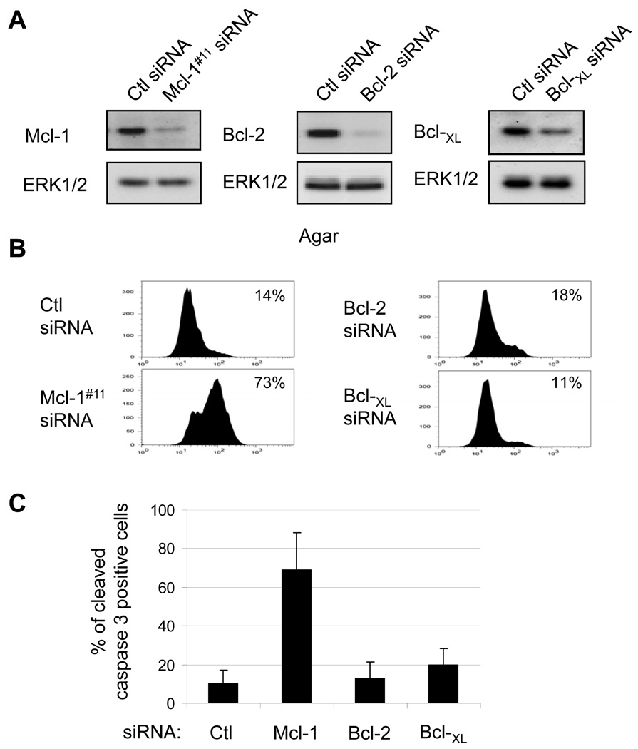
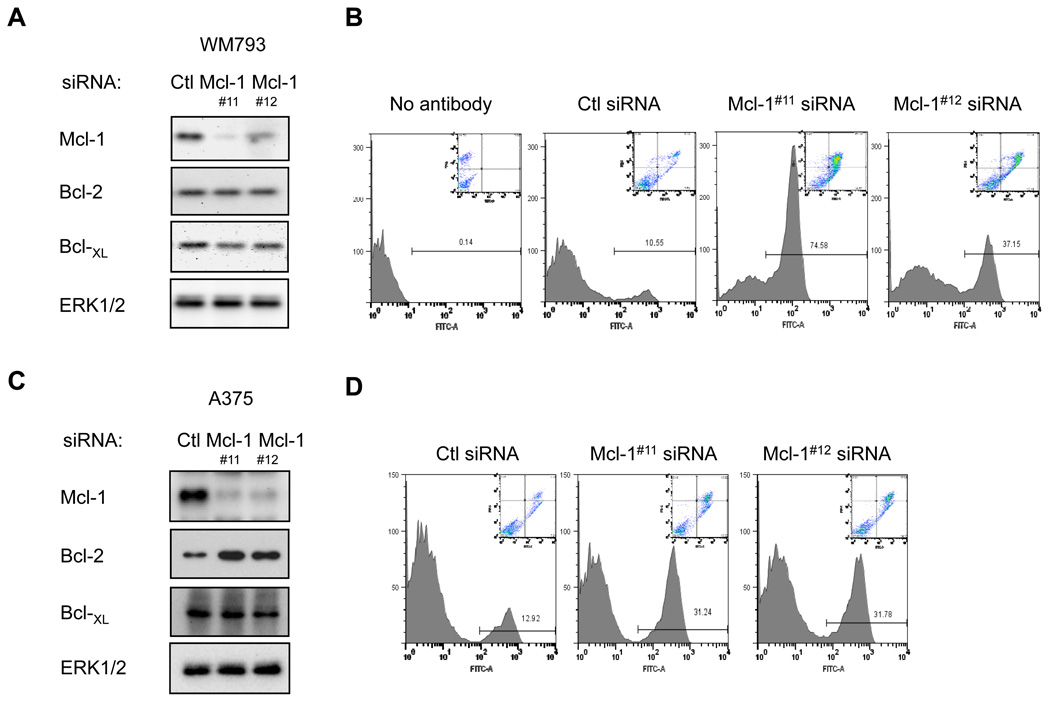
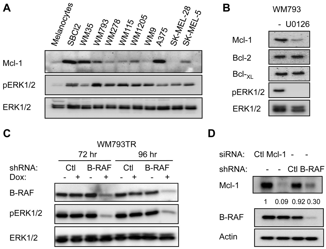
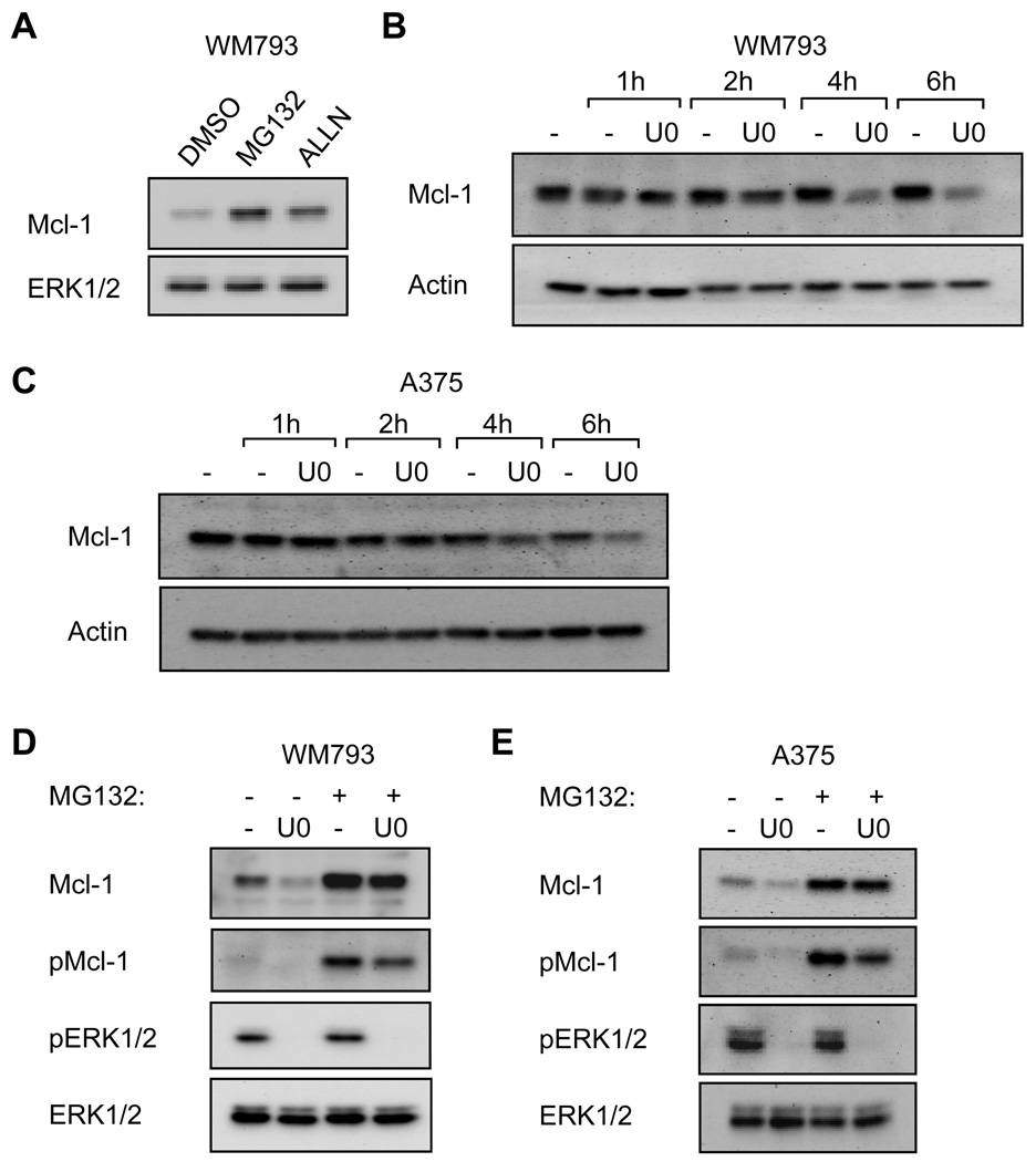
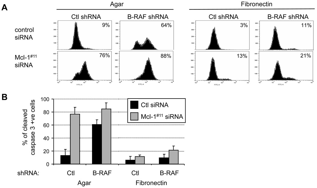
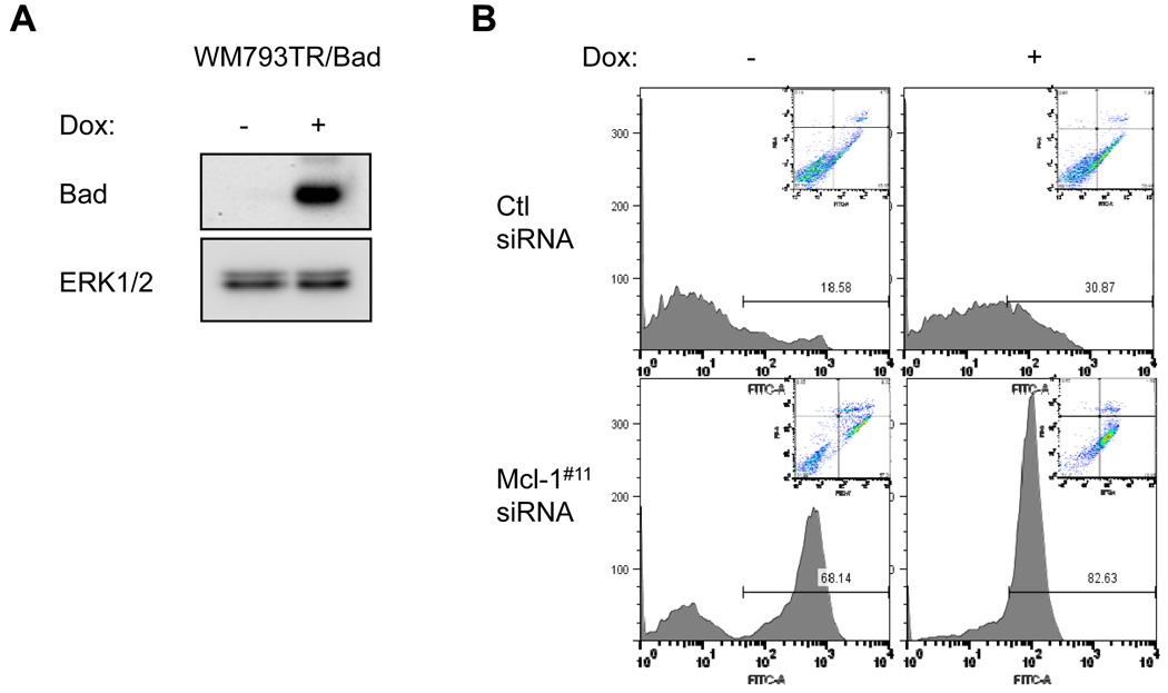
Melanoma is a particularly aggressive tumor type that exhibits a high level of resistance to apoptosis. The serine/threonine kinase B-RAF is mutated in 50% to 70% of melanomas and protects melanoma cells from anoikis, a form of apoptosis induced by lack of adhesion or adhesion to an inappropriate matrix. Mutant B-RAF down-regulates two BH3-only proapoptotic proteins, Bim(EL) and Bad. BH3-only proteins act, at least in part, by sequestering prosurvival Bcl-2 family proteins and preventing them from inhibiting the mitochondrial apoptotic pathway. Several Bcl-2 proteins are up-regulated in melanoma; however, the mechanisms of up-regulation and their role in melanoma resistance to anoikis remain unclear. Using RNA interference, we show that depletion of Mcl-1 renders mutant B-RAF melanoma cells sensitive to anoikis. By contrast, minor effects were observed following depletion of either Bcl-2 or Bcl-(XL). Mcl-1 expression is enhanced in melanoma cell lines compared with melanocytes and up-regulated by the B-RAF-MEK-extracellular signal-regulated kinase 1/2 pathway through control of Mcl-1 protein turnover. Similar to B-RAF knockdown cells, adhesion to fibronectin protected Mcl-1 knockdown cells from apoptosis. Finally, expression of Bad, which does not sequester Mcl-1, further augmented apoptosis in nonadherent Mcl-1 knockdown cells. Together, these data support the notion that BH3 mimetic compounds that target Mcl-1 may be effective for the treatment of melanoma in combinatorial strategies with agents that disrupt fibronectin-integrin signaling.


Conflict of interest statement

Disclosure of Potential Conflicts of Interest

No potential conflicts of interest were disclosed.

Figures

FIGURE 1
FIGURE 1
Mcl-1 knockdown promotes cleavage of caspase 3 in non-adherent melanoma cells. WM793 cells were transfected with control, Mcl-1, Bcl-2 or Bcl-XL siRNA, as indicated. (A) Seventy-two hours post-transfection, cell lysates were analyzed by Western blotting for Mcl-1, Bcl-2 or Bcl-XL. Total ERK1/2 was used as a loading control. (B) 72 hours following transfection, WM793 cells were serum starved for 24 hours and then replated onto agar-coated dishes in serum-free medium. After 48 hours, cells were analyzed for cleaved caspase 3 using flow cytometry. The fluorescence intensity is measured on the x axis and cell counts on the y-axis. The percentages of cells staining positive in each condition are indicated. (C) Quantitation of the data from (B) is presented as the average percentage of cells staining positive for cleaved caspase 3 from three independent experiments.
FIGURE 2
FIGURE 2
Mcl-1 knockdown sensitizes WM793 and A375 cells to anoikis. (A) WM793 cells were transfected with control, Mcl-1#11, or Mcl-1#12 siRNA. Cell lysates were analyzed by Western blotting for Mcl-1, Bcl-2, Bcl-XL and ERK1/2. (B) Following transfection and serum starvation, WM793 cells were replated onto agar-coated dishes for 48 hours in serum-free medium. Harvested cells were analyzed for annexin-V staining and propidium iodide uptake. The main trace shows annexin V-FITC staining versus relative cell number. Inset depicts annexin V-FITC staining versus PI incorporation. (C) and (D) As for A and B, except that A375 cells were utilized. Shown are representative traces from one of three independent experiments.
FIGURE 3
FIGURE 3
Mcl-1 is highly expressed in melanoma cells. (A) Protein cell lysates from human melanocytes (NHEM) and melanoma cells (Sbcl2, WM35, WM793, WM278, WM115, WM1205, WM9, A375, SK-MEL-28 and SK-MEL-5) were analyzed for Mcl-1, phospho-ERK1/2 and total ERK1/2 levels. (B) WM793 cells were treated with U0126 for 5 hours and lysates were analyzed by Western blotting. (C) WM793TR-Ctl shRNA and WM793TR-B-RAF shRNA cell lines were untreated or treated with 100 ng/ml doxycycline for 72 and 96 hours. Cell lysates were analyzed by Western blotting for B-RAF, phospho-ERK1/2, and total ERK1/2. (D) Ctl and Mcl-1 knockdown WM793 cells, and WM793TR-Ctl shRNA and WM793TR-B-RAF shRNA treated with doxycycline for 96 hours were analyzed by Western blotting for levels of Mcl-1, B-RAF and actin. The levels of Mcl-1 normalized to actin are indicated for each sample.
FIGURE 4
FIGURE 4
Mcl-1 protein turnover is regulated by B-RAF-MEK signaling. (A) WM793 cells were treated with 10 µM MG132 or 50 µM ALLN for 5 hours. Cell lysates were analyzed by Western blotting. (B) WM793 cells were treated with 10 µg/ml cycloheximide for 1 hour after which one culture dish was taken as the time 0. Cells were then treated with either DMSO or 5 µM U0126 for the indicated time. Cell lysates were analyzed by Western blotting for Mcl-1 and actin (loading control). (C) As for B, except A375 cells were used. (D) WM793 cells were treated for 6 hours with 10 µM MG132 and/or 5 µM U0126, as indicated. Cell lysates were analyzed by Western blotting for levels of total and phospho-Mcl-1, and total and phospho-ERK1/2. (E) As for D, except that A375 cells were utilized. Shown are representative blots from one of three independent experiments.
FIGURE 5
FIGURE 5
Adhesion to fibronectin protects Mcl-1 knockdown cells from apoptosis. (A) Doxycycline-treated WM793TR-Ctl shRNA and WM793TR-B-RAF shRNA cells were transfected with either Ctl or Mcl-1 siRNAs. Following transfection and serum starvation, WM793 cells were replated onto agar or fibronectin-coated dishes for 48 hours in serum-free medium. Harvested cells were analyzed for cleaved caspase 3 staining by flow cytometry. (B) Quantitation of cleaved caspase 3 staining from three independent experiments.
FIGURE 6
FIGURE 6
Bad expression augments sensitivity to anoikis in Mcl-1 knockdown cells. (A) WM793TR-Bad cells were treated for 72 hrs with 100 ng/ml doxycycline. Lysates were analyzed by Western blotting for Bad and ERK1/2, as a loading control. (B) WM793TR-Bad cells were transfected with Ctl or Mcl-1 siRNA and used in anoikis assays. Harvested cells were analyzed for annexin-V staining and propidium iodide uptake. Shown are representative results from one of three independent experiments.

Similar articles

Cited by

References

    1. Frisch SM, Screaton RA. Anoikis mechanisms. Curr Opin Cell Biol. 2001;13:555–562. - PubMed
    1. Adams JM, Cory S. The Bcl-2 apoptotic switch in cancer development and therapy. Oncogene. 2007;26:1324–1337. - PMC - PubMed
    1. Letai A, Bassik MC, Walensky LD, et al. Distinct BH3 domains either sensitize or activate mitochondrial apoptosis, serving as prototype cancer therapeutics. Cancer Cell. 2002;2:183–192. - PubMed
    1. Kuwana T, Bouchier-Hayes L, Chipuk JE, et al. BH3 domains of BH3-only proteins differentially regulate Bax-mediated mitochondrial membrane permeabilization both directly and indirectly. Mol Cell. 2005;17:525–535. - PubMed
    1. Willis SN, Fletcher JI, Kaufmann T, et al. Apoptosis initiated when BH3 ligands engage multiple Bcl-2 homologs, not Bax or Bak. Science. 2007;315:856–859. - PubMed

Publication types

MeSH terms

Substances