. 2009 Jun 12;284(24):16513-16521.
doi: 10.1074/jbc.M109.001875. Epub 2009 Apr 16.

Identification and characterization of the Na+/H+ antiporter Nhas3 from the thylakoid membrane of Synechocystis sp. PCC 6803

Affiliations

Identification and characterization of the Na+/H+ antiporter Nhas3 from the thylakoid membrane of Synechocystis sp. PCC 6803

Kenta Tsunekawa et al. J Biol Chem. .

Abstract

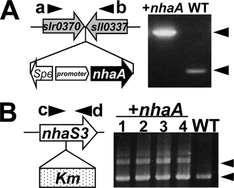
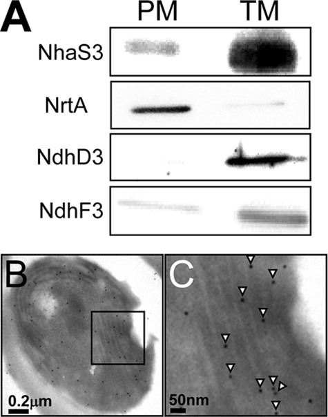
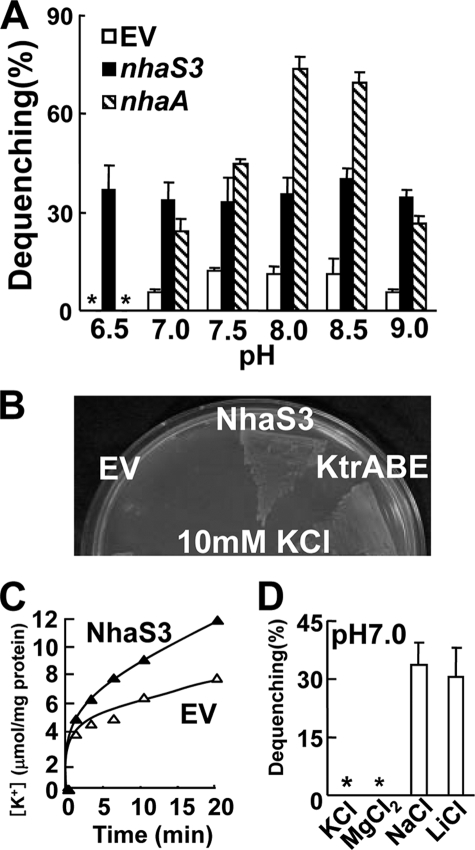
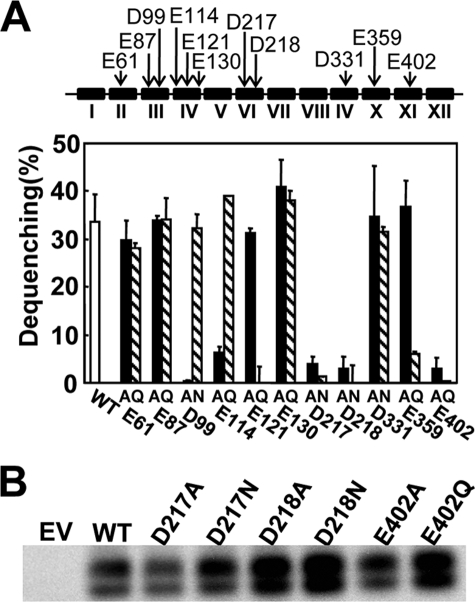
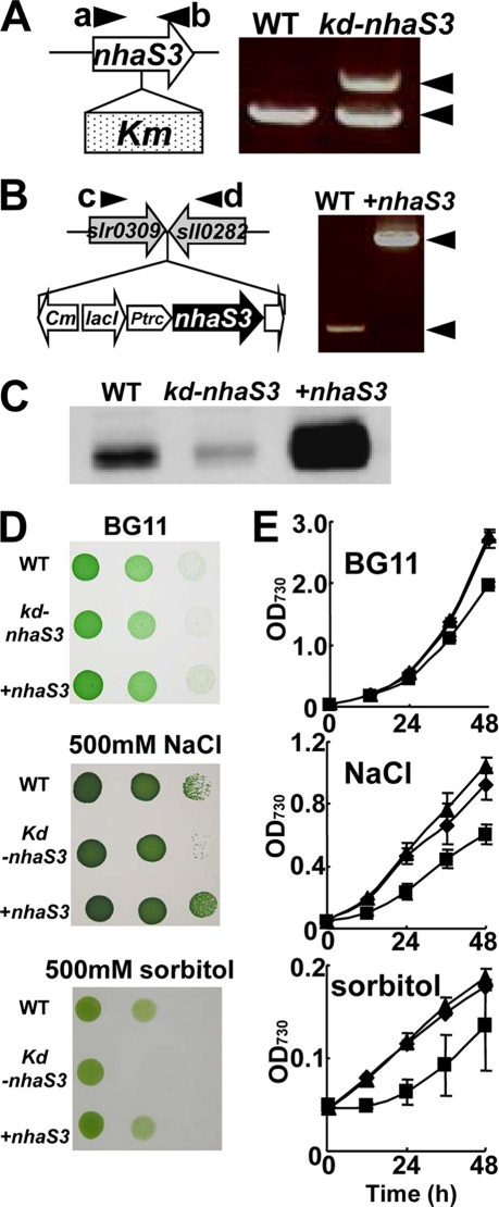
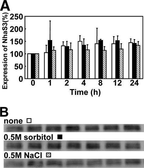
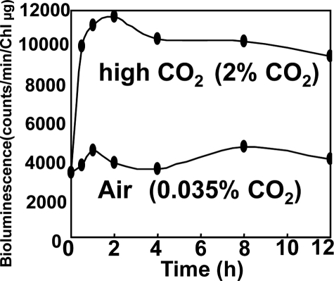
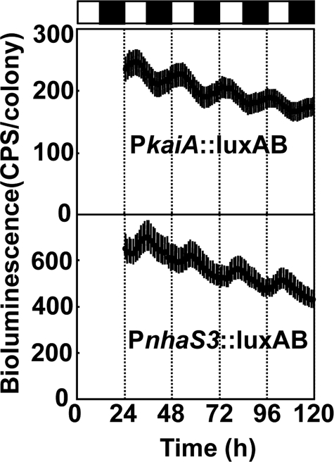
Na+/H+ antiporters influence proton or sodium motive force across the membrane. Synechocystis sp. PCC 6803 has six genes encoding Na+/H+ antiporters, nhaS1-5 and sll0556. In this study, the function of NhaS3 was examined. NhaS3 was essential for growth of Synechocystis, and loss of nhaS3 was not complemented by expression of the Escherichia coli Na+/H+ antiporter NhaA. Membrane fractionation followed by immunoblotting as well as immunogold labeling revealed that NhaS3 was localized in the thylakoid membrane of Synechocystis. NhaS3 was shown to be functional over a pH range from pH 6.5 to 9.0 when expressed in E. coli. A reduction in the copy number of nhaS3 in the Synechocystis genome rendered the cells more sensitive to high Na+ concentrations. NhaS3 had no K+/H+ exchange activity itself but enhanced K+ uptake from the medium when expressed in an E. coli potassium uptake mutant. Expression of nhaS3 increased after shifting from low CO2 to high CO2 conditions. Expression of nhaS3 was also found to be controlled by the circadian rhythm. Gene expression peaked at the beginning of subjective night. This coincided with the time of the lowest rate of CO2 consumption caused by the ceasing of O2-evolving photosynthesis. This is the first report of a Na+/H+ antiporter localized in thylakoid membrane. Our results suggested a role of NhaS3 in the maintenance of ion homeostasis of H+, Na+, and K+ in supporting the conversion of photosynthetic products and in the supply of energy in the dark.


Figures

FIGURE 1.
FIGURE 1.
Introduction of E. coli nhaA into Synechocystis. A, E. coli nhaA under control of the iron-inducible promoter was introduced at a site between slr0370 and sll0337 of Synechocystis chromosome (left panel). During growth in BG11 medium, nhaA is constitutively expressed. Correct insertion of the expression construct was confirmed by PCR on genomic DNA using specific primers a and b; the results are shown in the right panel. WT, wild type; +nhaA, nhaA-expressing cells; Spe, spectinomycin resistance gene. B, disruption of nhaS3 in the strain expressing E. coli nhaA was performed by insertion of the kanamycin resistance gene (Km) into nhaS3. Correct integration of the kanamycin gene was tested by PCR on genomic DNA using specific primers c and d. The results for four independent clones and WT are shown in the right panel. Note that nhaA could not replace nhaS3.
FIGURE 2.
FIGURE 2.
Membrane localization of NhaS3. A, membrane fractions were prepared by aqueous polymer two-phase partitioning and sucrose density gradient and separated by SDS-PAGE. NhaS3, NrtA, NdhD3, and NdhF3 proteins were detected on Western blots using the corresponding antibodies. PM, plasma membrane; TM, thylakoid membrane. B and C, cross-section of a Synechocystis cell immunolabeled using an anti-NhaS3 antibody. C is an enlarged section of B. NhaS3 protein, indicated by the presence of gold particles (arrowheads in C), was detected in the thylakoid membrane.
FIGURE 3.
FIGURE 3.
Transport activity of NhaS3. A, pH dependence of Na+/H+ antiporter activities of NhaS3 in inverted membrane vesicles prepared from nhaS3-expressing E. coli. Fluorescence from acridine orange was monitored. The percentage of fluorescence dequenching observed after the addition of 5 mm NaCl was plotted relative to that of lactate-induced quenching. EV, empty vector; *, not detected. B, complementation of an E. coli K+ uptake mutant (LB2003) by NhaS3. Growth of E. coli LB2003 expressing nhaS3 or Synechocystis K+ uptake transporter KtrABE on synthetic solid medium containing 10 mm KCl. C, K+ uptake activity by NhaS3 in E. coli. Cells containing NhaS3 or the empty vector were incubated with 1 mm KCl in HEPES-NaOH, pH 7.5, and aliquots were withdrawn at the indicated times. D, measurement of K+ or Mg2+ efflux mediated by NhaS3. K+, Mg2+, or Li+, instead of Na+ were added to membrane vesicles prepared from nhaS3-expressing E. coli cells. Antiporter activity was determined by the same procedure as in A. *, not detected.
FIGURE 4.
FIGURE 4.
Na+/H+ antiporter activities in the inverted membrane vesicles of E. coli expressing wild type NhaS3 or NhaS3 variants. A, antiporter activities of the NhaS3 variants. The locations of the negatively charged residues (Asp or Glu) that were replaced with Ala or Asn/Gln are indicated with arrows (top). The Roman numerals designate the numbers of the putative transmembrane spanning domains predicted by the TopPredII algorithm and counted from the N terminus of NhaS3. Na+/H+ antiporter activity was determined in inverted membrane vesicles by measuring the percentage of fluorescence dequenching observed after addition of 5 mm NaCl (bottom). B, immunological detection of NhaS3 variants containing mutations at Asp217, Asp218, and Glu402 that showed no transport activities in A. The membrane proteins isolated from the strains expressing the nhaS3 variants were separated by SDS-polyacrylamide gel electrophoresis (12% acrylamide) and blotted, and the blots were probed using anti-NhaS3 antibodies. WT, wild type; EV, empty vector.
FIGURE 5.
FIGURE 5.
Inhibition of growth of nhaS3 knockdown Synechocystis cells by salt stress and hyperosmotic stress. A, creation of a nhaS3 knockdown strain (kd-nhaS3). The nhaS3 gene was disrupted in Synechocystis by insertion of a kanamycin resistance gene (Km). Integration of Km was detected by PCR using primers a and b. Note that nhdS3 was not completely disrupted. B, overexpression of nhaS3 (+nhaS3). A construct containing the lacIq, Ptrc::nhaS3, and chloramphenicol resistance (Cm) gene was integrated into the TS4 sites (between slr0309 and sll0282) in the Synechocystis genome. Integration of the overexpression construct was verified by PCR using primers c and d. C, validation of both the knockdown strain (kd-nhaS3) and the overexpression strain (+nhaS3). NhaS3 protein levels were detected by Western blot analysis. D, growth of the wild type (WT), nhaS3 knockdown strain (kd-nhaS3), and nhaS3 overexpression strain (+nhaS3) on solid BG11 medium or on BG11 medium containing either 500 mm NaCl or 500 mm sorbitol. E, growth test performed in liquid culture using the same strains and conditions as in D. The OD730 was measured at the times indicated. WT, circle; kd-nhaS3, square; +nhaS3, triangle.
FIGURE 6.
FIGURE 6.
Effect of salt stress or hyperosmotic stress on the expression of NhaS3. A, wild type cells cultured in liquid BG11 medium (OD730 = 0.5) were transferred to the same medium (white bars) or to hyperosmotic medium containing either 500 mm NaCl (black bars) or 500 mm sorbitol (hatched bars) and cultured for the time indicated. B, cells samples taken from the cultures in A at the times indicated were subjected to Western blot analysis using anti-NhaS3 antibodies.
FIGURE 7.
FIGURE 7.
Induction of nhaS3 expression under high CO2 conditions. Synechocystis cells containing a luciferase reporter gene under control of the nhaS3 promoter (PnhaS3::luxAB) were grown under low CO2 conditions (0.035% CO2 in air) and transferred to high CO2 conditions (2% CO2 in air). Control cells were left at low CO2 conditions (0.035% in air). The time scale represents the time after transfer. At the times indicated the luciferase activity was determined.
FIGURE 8.
FIGURE 8.
Circadian rhythm of nhaS3 expression. Synechocystis cells containing a luciferase reporter gene under control of the nhaS3 promoter (PnhaS3::luxAB) were entrained by a 12-h dark period and then shifted to continuous light. The PkaiA::luxAB reporter strain, which shows circadian bioluminescence rhythms,4 was used as control. The time scale represents the actual time after transfer to continuous light conditions. The bioluminescence was determined every hour. Each point indicates the average ± standard deviation from seven replicates. The white and black boxes above the graphs represent subjective day and night, respectively.

Similar articles

Cited by

References

    1. Padan E., Schuldiner S. ( 1994) Biochim. Biophys. Acta 1185, 129– 151 - PubMed
    1. Padan E., Schuldiner S. ( 1994) Biochim. Biophys. Acta 1187, 206– 210 - PubMed
    1. Hunte C., Screpanti E., Venturi M., Rimon A., Padan E., Michel H. ( 2005) Nature 435, 1197– 1202 - PubMed
    1. Ohyama T., Igarashi K., Kobayashi H. ( 1994) J. Bacteriol. 176, 4311– 4315 - PMC - PubMed
    1. Hamada A., Hibino T., Nakamura T., Takabe T. ( 2001) Plant Physiol. 125, 437– 446 - PMC - PubMed

Publication types

LinkOut - more resources