. 2009;4(5):662-73.
doi: 10.1038/nprot.2009.33.

A protocol for the culture and differentiation of highly polarized human retinal pigment epithelial cells

Affiliations

A protocol for the culture and differentiation of highly polarized human retinal pigment epithelial cells

Shozo Sonoda et al. Nat Protoc. 2009.

Abstract

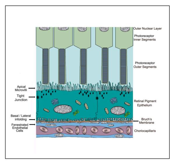
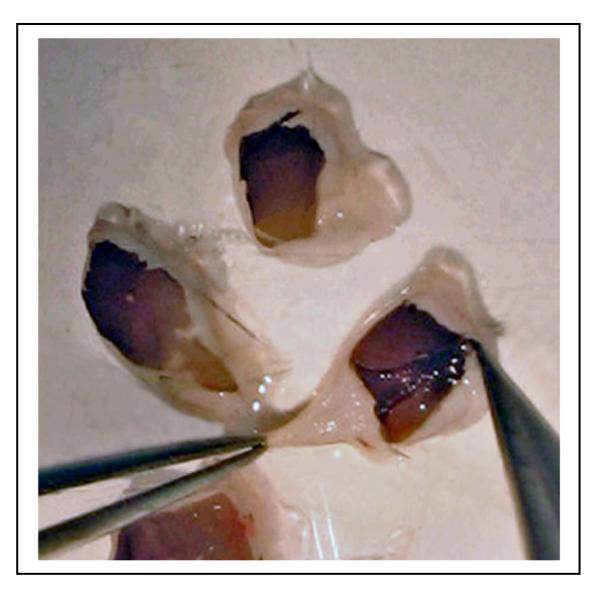
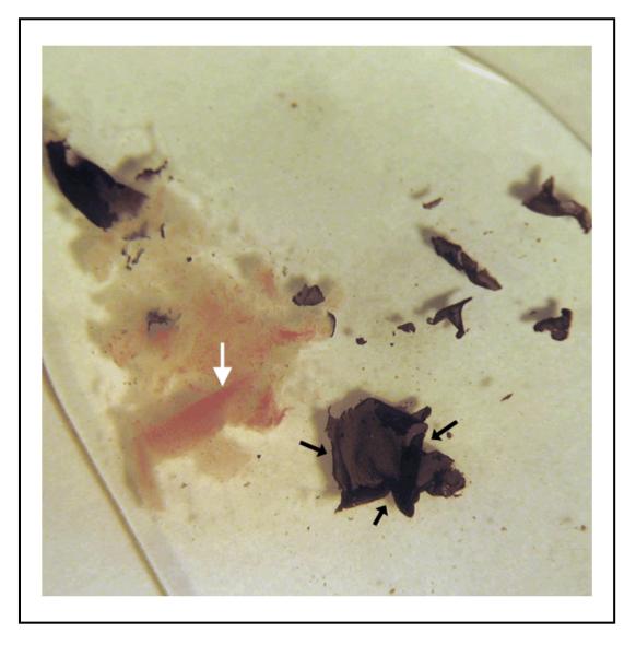
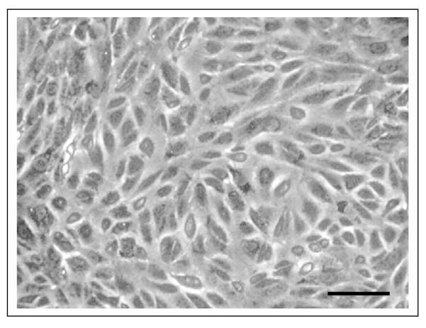
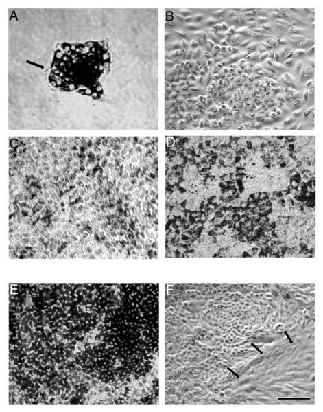
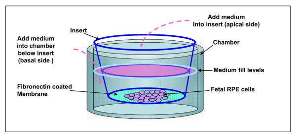
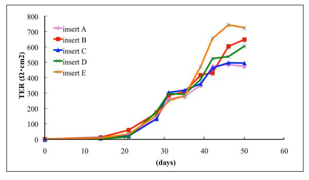
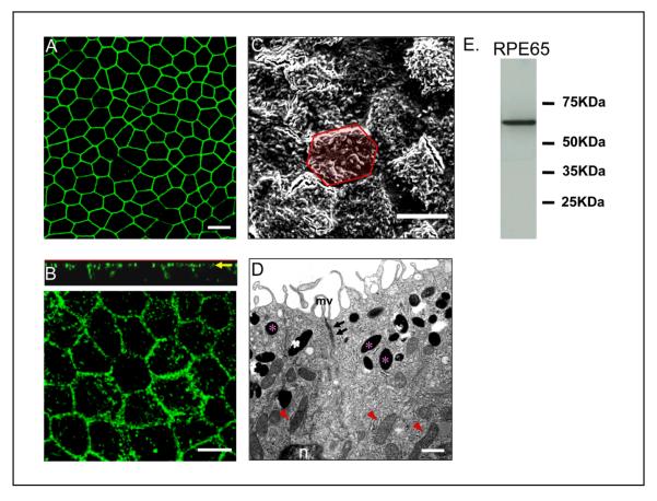
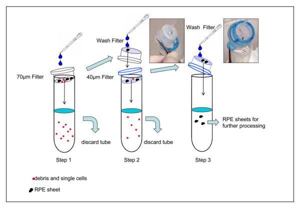
We provide our detailed, standardized in vitro protocol for the culture and differentiation of human retinal pigment epithelial (RPE) cells into a highly polarized and functional monolayer. Disruption of the polarized RPE function plays an important role in the pathogenesis of common blinding disorders of the retina. The availability of this polarized RPE monolayer allows for reproducible evaluation of RPE function, modeling of RPE dysfunction in retinal disease and in vitro evaluation of new therapies. The protocol, which takes approximately 6 weeks to complete, describes the culture of RPE from human fetal donor eyes, the differentiation of these cells into a polarized monolayer with high transepithelial resistance and morphologic characteristics that mimic the RPE monolayer in vivo. By modifying the procedure for initial isolation of pure RPE cells and the culture conditions used in existing protocols, we have established a standardized protocol that provides highly reproducible RPE monolayers from the same donor eye.


Figures

Figure 1
Figure 1
Diagram of the outer retina illustrating the polarized nature of the RPE monolayer and its relationship to the photoreceptor inner and outer segments, Bruch’s membrane and the choriocapillaris. The RPE shows apical microvilli, tight junctions and basal/lateral infolding, The endothelial cells of the choroid are fenestrated.
Figure 2
Figure 2
Human fetal eye with the first incision. A radial incision was made into the cornea (arrow) using a sterile Teflon coated razor blade without high pressure. Bar equals 5mm.
Figure 3
Figure 3
The fetal eye is split into cornea/iris (anterior segment), lens, and posterior eye cup. When the cornea/ iris complex is removed, the lens easily comes off with the anterior segment. Bar equals 5 mm.
Figure 4
Figure 4
The human fetal eye is dissected into 4 quadrants. An incision should be made as close to the optic disc with a razor blade in order for the eye cup to lay flat (a). The sensory retina (asterisk) is firmly attached to the optic disc. The sensory retina is gently removed from the RPE layer with a sterile disposable inoculating loop, care being taken not to make scratches into the RPE layer (b). After removal of retina, there is no obvious damage to the RPE layer (c).
Figure 5
Figure 5
The RPE/choroid layer is peeled off from the sclera using forceps. Usually the layer can be isolated as an intact sheet.
Figure 6
Figure 6
Intact RPE sheets (black arrows) are peeled from the choroid with fine forceps under a dissecting microscope. Vessels (white arrow) in choroid are clearly visible after removing the RPE layer.
Figure 7
Figure 7
Light microscopic images of early passage confluent fetal RPE cells grown on a plastic tissue culture plate. Cells do not show hexagonal shape and lack pigmentation under these culture conditions. Bar equals 40 μm.
Figure 8
Figure 8
Light microscopic images showing pattern of time dependent growth of normal RPE in a T75 flask (a-d), and highly pigmented (e) and overgrown multi-layers of RPE (f) for comparison. (a) Day1; Cells are attached and initial growth of the culture occurred in the form of small islands, consisting of 15-25 RPE cells. (b) Day4; Rapid growth of cells with a decrease in pigment is seen. (c) Day14; Cells show evidence of repigmentation. (d) Day21; Cells showing hexagonal shape and increase in pigment density. (e) Hyperpigmented RPE on day 21. These cell are not suitable for establishing polarity. (f) RPE cells with abnormal morphology on day 21. Arrows show cells with fibroblastic morphology. Bar equals 60 μm.
Figure 9
Figure 9
Schematic diagram showing culture of highly differentiated, polarized human RPE in culture wells with a Transwell insert.
Figure 10
Figure 10
Time dependent increase in TER from multiple Transwells containing RPE from a single donor. Note the time-dependent increase in TER beginning after 15 days in culture, and that RPE from multiple Transwells (inserts A-E) develop TER in a reproducible manner.
Figure 11
Figure 11
Evidence for differentiation and polarization in human fetal RPE cells cultured on Transwell filters for 4 weeks. (a) Immunofluorescence pattern of tight junction protein ZO-1 visualized by confocal microscopy. (b) Localization of Na/K ATPase to the apical plasma membrane as lower panel and vertical (X-Z) section (upper panel). Na/K ATPase is preferentially located at the apical surface. (c) Well differentiated apical microvilli observed by scanning electron microscopy. This image contains 17 cells; the yellow hexagonal area in the figure outlines a single RPE cell. (d) Localization of pigment in the apical cytoplasm (asterisk), nucleus (n) in the basal side of the cell, apical tight-junctional complexes (arrows) and mitochondria (arrowheads) by transmission electron microscopy. (e) The expression of the RPE-specific protein, RPE65 was confirmed by western blot analysis in lysate from 4week old polarized RPE cells. a,b,c; Bar equals 10 μm. d; Bar equals 1μm.
Figure 12
Figure 12
Steps involved in the isolation of pure RPE sheets. Please note two different filters (70 and 40 μm) are used. Holding buffer (25 ml) was used for filtration in each step. Red dots indicate debris and single cells, whereas black dots represent RPE sheets.
Figure 13
Figure 13
Measurment of TER of polarized human RPE using a voltohmmeter. All TER measurements are made in a cell culture hood within 3 min of removal of Transwells from the incubator. The lower end of the longer electrode (inset asterisk) is inserted into the basolateral medium and shorter electrode into the apical medium.

References

    1. Thumann G, Hoffmann S, Hinton DR. Cell biology of the retinal pigment epithelium. In: Ryan SJ, editor. Retina. 4rd ed Vol. 1. Elsevier Mosby; Philadelphia, USA: 2006. pp. 137–152.
    1. Rodriguez-Boulan E, Nelson WJ. Morphogenesis of the polarized epithelial cell phenotype. Science. 1989;245:718–725. - PubMed
    1. Marmor MF, Abdul-Rahim AS, Cohen DS. The effect of metabolic inhibitors on retinal adhesion and subretinal fluid resorption. Invest Ophthalmol Vis Sci. 1980;19:893–903. - PubMed
    1. Miller SS, Hughes BA, Machen TE. Fluid transport across retinal pigment epithelium is inhibited by cyclic AMP. Proc Natl Acad Sci U S A. 1982;79:2111–2115. - PMC - PubMed
    1. Anderson JM, Van Itallie CM. Tight junctions and the molecular basis for regulation of paracellular permeability. Am J Physiol. 1995;269:G467–475. - PubMed

Publication types