Structural basis of N-end rule substrate recognition in Escherichia coli by the ClpAP adaptor protein ClpS
- PMID: 19373253
- PMCID: PMC2680879
- DOI: 10.1038/embor.2009.62
Structural basis of N-end rule substrate recognition in Escherichia coli by the ClpAP adaptor protein ClpS
Erratum in
- EMBO Rep. 2009 Jun;10(6):662
Abstract
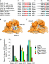
In Escherichia coli, the ClpAP protease, together with the adaptor protein ClpS, is responsible for the degradation of proteins bearing an amino-terminal destabilizing amino acid (N-degron). Here, we determined the three-dimensional structures of ClpS in complex with three peptides, each having a different destabilizing residue--Leu, Phe or Trp--at its N terminus. All peptides, regardless of the identity of their N-terminal residue, are bound in a surface pocket on ClpS in a stereo-specific manner. Several highly conserved residues in this binding pocket interact directly with the backbone of the N-degron peptide and hence are crucial for the binding of all N-degrons. By contrast, two hydrophobic residues define the volume of the binding pocket and influence the specificity of ClpS. Taken together, our data suggest that ClpS has been optimized for the binding and delivery of N-degrons containing an N-terminal Phe or Leu.
Conflict of interest statement
The authors declare no conflict of interest.
Figures


Similar articles
-
ClpS is an essential component of the N-end rule pathway in Escherichia coli.Nature. 2006 Feb 9;439(7077):753-6. doi: 10.1038/nature04412. Nature. 2006. PMID: 16467841
-
The ClpS adaptor mediates staged delivery of N-end rule substrates to the AAA+ ClpAP protease.Mol Cell. 2011 Jul 22;43(2):217-28. doi: 10.1016/j.molcel.2011.06.009. Mol Cell. 2011. PMID: 21777811 Free PMC article.
-
Distinct structural elements of the adaptor ClpS are required for regulating degradation by ClpAP.Nat Struct Mol Biol. 2008 Mar;15(3):288-94. doi: 10.1038/nsmb.1392. Epub 2008 Feb 24. Nat Struct Mol Biol. 2008. PMID: 18297088
-
The N-end rule pathway for regulated proteolysis: prokaryotic and eukaryotic strategies.Trends Cell Biol. 2007 Apr;17(4):165-72. doi: 10.1016/j.tcb.2007.02.001. Epub 2007 Feb 15. Trends Cell Biol. 2007. PMID: 17306546 Review.
-
Proteolysis: Adaptor, adaptor, catch me a catch.Curr Biol. 2004 Nov 9;14(21):R924-6. doi: 10.1016/j.cub.2004.10.015. Curr Biol. 2004. PMID: 15530384 Review.
Cited by
-
Insights into degradation mechanism of N-end rule substrates by p62/SQSTM1 autophagy adapter.Nat Commun. 2018 Aug 17;9(1):3291. doi: 10.1038/s41467-018-05825-x. Nat Commun. 2018. PMID: 30120248 Free PMC article.
-
ClpP protease activation results from the reorganization of the electrostatic interaction networks at the entrance pores.Commun Biol. 2019 Nov 13;2:410. doi: 10.1038/s42003-019-0656-3. eCollection 2019. Commun Biol. 2019. PMID: 31754640 Free PMC article.
-
A pangenomic analysis of the Nannochloropsis organellar genomes reveals novel genetic variations in key metabolic genes.BMC Genomics. 2014 Mar 19;15:212. doi: 10.1186/1471-2164-15-212. BMC Genomics. 2014. PMID: 24646409 Free PMC article.
-
Signaling Pathways Regulated by UBR Box-Containing E3 Ligases.Int J Mol Sci. 2021 Aug 3;22(15):8323. doi: 10.3390/ijms22158323. Int J Mol Sci. 2021. PMID: 34361089 Free PMC article. Review.
-
Structural basis for the N-degron specificity of ClpS1 from Arabidopsis thaliana.Protein Sci. 2021 Mar;30(3):700-708. doi: 10.1002/pro.4018. Epub 2020 Dec 30. Protein Sci. 2021. PMID: 33368743 Free PMC article.
References
-
- Dougan DA, Reid BG, Horwich AL, Bukau B (2002) ClpS, a substrate modulator of the ClpAP machine. Mol Cell 9: 673–683 - PubMed
-
- Emsley P, Cowtan K (2004) Coot: model-building tools for molecular graphics. Acta Crystallogr D Biol Crystallogr 60: 2126–2132 - PubMed
-
- Erbse A, Schmidt R, Bornemann T, Schneider-Mergener J, Mogk A, Zahn R, Dougan DA, Bukau B (2006) ClpS is an essential component of the N-end rule pathway in Escherichia coli. Nature 439: 753–756 - PubMed
Publication types
MeSH terms
Substances
LinkOut - more resources
Full Text Sources
Other Literature Sources
Molecular Biology Databases
Miscellaneous

