. 2009 May;10(5):466-73.
doi: 10.1038/embor.2009.55. Epub 2009 Apr 17.

Molecular discrimination of structurally equivalent Lys 63-linked and linear polyubiquitin chains

Affiliations

Molecular discrimination of structurally equivalent Lys 63-linked and linear polyubiquitin chains

David Komander et al. EMBO Rep. 2009 May.

Abstract

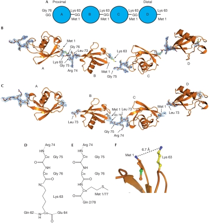
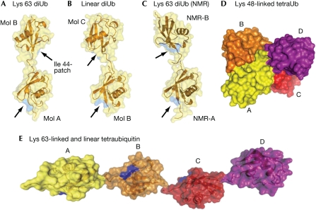
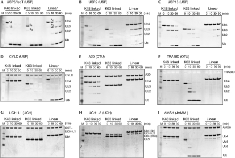
At least eight types of ubiquitin chain exist, and individual linkages affect distinct cellular processes. The only distinguishing feature of differently linked ubiquitin chains is their structure, as polymers of the same unit are chemically identical. Here, we have crystallized Lys 63-linked and linear ubiquitin dimers, revealing that both adopt equivalent open conformations, forming no contacts between ubiquitin molecules and thereby differing significantly from Lys 48-linked ubiquitin chains. We also examined the specificity of various deubiquitinases (DUBs) and ubiquitin-binding domains (UBDs). All analysed DUBs, except CYLD, cleave linear chains less efficiently compared with other chain types, or not at all. Likewise, UBDs can show chain specificity, and are able to select distinct linkages from a ubiquitin chain mixture. We found that the UBAN (ubiquitin binding in ABIN and NEMO) motif of NEMO (NF-kappaB essential modifier) binds to linear chains exclusively, whereas the NZF (Npl4 zinc finger) domain of TAB2 (TAK1 binding protein 2) is Lys 63 specific. Our results highlight remarkable specificity determinants within the ubiquitin system.


Conflict of interest statement

The authors declare that they have no conflict of interest.

Figures

Figure 1
Figure 1
Structure of Lys 63 and linear ubiquitin chains. (A) Nomenclature for polyubiquitin chains. The proximal molecule is linked through its carboxy terminus to a substrate lysine residue, or has a free carboxy-terminal diGly (GG) motif in unattached chains. (B,C) Four equivalent ubiquitin molecules, corresponding to two adjacent asymmetric units within the crystal lattice, are shown in cartoon representation. 2∣Fo∣−∣Fc∣ electron density at 1σ is drawn for the linkage residues between molecules A–B and C–D for Lys 63-linked diubiquitin, and for B–C in linear diubiquitin. (D) Chemical representation of the Lys 63 linkage. Other isopeptide linkages (for example, Lys 48 linkages) differ only in the type of neighbouring residues. (E) Representation of the peptide linkage in a linear ubiquitin chain between Gly 76 and Met 1 of the second molecule. (F) Close spatial location of Lys 63 and Met 1 (distance of 6.7 Å) allow similar conformation of linear and Lys 63-linked chains. Ub, ubiquitin.
Figure 2
Figure 2
Similarities and differences between differentially linked ubiquitin polymers. (AC) A semitransparent surface covers the ubiquitin molecules in cartoon representation, and the position of the hydrophobic surface patch formed by Ile 44-Val 70-Leu 8 is shown in blue on the surface, indicated by arrows. The diubiquitin molecules are aligned on the proximal ubiquitin moiety. (A) Structure of Lys 63-linked diubiquitin. (B) Diubiquitin orientations derived from the linear diubiquitin crystal structure (representing mol B/mol C in the Lys 63 structure). (C) Diubiquitin orientation derived from NMR analysis (Varadan et al, 2004; coordinates were kindly provided by D. Fushman). (D) Model of Lys 48-linked tetraubiquitin (pdb-id 2o6v; Eddins et al, 2007). (E) Model of linear or Lys 63-linked tetraubiquitin. NMR, nuclear magnetic resonance; Ub, ubiquitin.
Figure 3
Figure 3
Specificity of deubiquitinating enzymes. Time course analysis of degradation of tetraubiquitin by different DUBs was visualized by silver staining and performed as described by Komander et al (2008). (A) IsoT/USP5, (B) USP2, (C) USP15, (D) CYLD, (E) A20, (F) TRABID, (G) UCH-L1, (H) UCH-L3 and (I) AMSH. Polyubiquitin chains (n>2) run at different sizes on SDS–PAGE, labelled in (A): *4K48-Ub4, *3K48-Ub3, $4K63-Ub4, $3K63-Ub3, #4linear Ub4, #3linear Ub3. AMSH, associated molecule with the SH3 domain of STAM; DUB, deubiquitinase; GST, glutathione S-transferase; JAMM, JAB1/MPN/Mov34; M, marker; OTU, ovarian tumour; SDS–PAGE, SDS–polyacrylamide gel electrophoresis; Ub, ubiquitin; UCH, ubiquitin carboxy-terminal hydrolase; USP, ubiquitin specific protease.
Figure 4
Figure 4
Binding of ubiquitin tetramers to selected ubiquitin-binding domains. (A) Pull-down analysis with immobilized GST-tagged UBDs incubated with 1.5 μg tetraubiquitin of different linkages. Three lanes per linkage correspond to 5% of input tetraubiquitin, GST–UBD-bound tetraubiquitin and GST control-bound tetraubiquitin. Ponceau-stained membranes are shown in supplementary Fig 3 online. (B) Pull-down analysis as in (A), in which the different tetraubiquitins were mixed and used as inputs. The Ponceau-stained membrane shows the GST–UBDs. (C) The input samples from (B; first four lanes) were silver and Coomassie stained, which indicated the antibody does not recognize different linkages equivalently. (D) Potential mechanisms of UBD binding to differently linked polyubiquitin chains. (E) Summary of specificity for all proteins tested. Full-length TRABID is further analysed in supplementary Fig 4 online. ABIN2(FL), A2O binding inhibitor of NF-κB signalling 2 (full length); AMSH, associated molecule with the SH3 domain of STAM; CARD, caspase recruitment domain; cIAP1, cellular inhibitor of apoptosis 1; DUBs, deubiquitinases; GST, glutathione S-transferase; IB, immunoblotting; JAMM, JAB1/MPN/Mov34; MUD1, UBA domain-contining protein mud1; NZF, Npl4 zinc finger; OTU, ovarian tumour; TAB2, TAK1 binding protein 2; Ub, ubiquitin; UBA, ubiquitin associated; UBAN, ubiquitin binding in ABIN and NEMO; UBD, ubiquitin-binding domain; UCH, ubiquitin carboxy-terminal hydrolase; USP, ubiquitin specific protease.

References

    1. Adhikari A, Xu M, Chen ZJ (2007) Ubiquitin-mediated activation of TAK1 and IKK. Oncogene 26: 3214–3226 - PubMed
    1. Clague MJ, Urbe S (2006) Endocytosis: the DUB version. Trends Cell Biol 16: 551–559 - PubMed
    1. Ea CK, Deng L, Xia ZP, Pineda G, Chen ZJ (2006) Activation of IKK by TNFα requires site-specific ubiquitination of RIP1 and polyubiquitin binding by NEMO. Mol Cell 22: 245–257 - PubMed
    1. Eddins MJ, Varadan R, Fushman D, Pickart CM, Wolberger C (2007) Crystal structure and solution NMR studies of Lys 48-linked tetraubiquitin at neutral pH. J Mol Biol 367: 204–211 - PubMed
    1. Gyrd-Hansen M et al. (2008) IAPs contain an evolutionarily conserved ubiquitin-binding domain that regulates NF-κB as well as cell survival and oncogenesis. Nat Cell Biol 10: 1309–1317 - PMC - PubMed

Publication types