

Pineal gland tumors: experience from the SEER database
- PMID: 19373436
- PMCID: PMC2804886
- DOI: 10.1007/s11060-009-9881-9
Pineal gland tumors: experience from the SEER database
Abstract
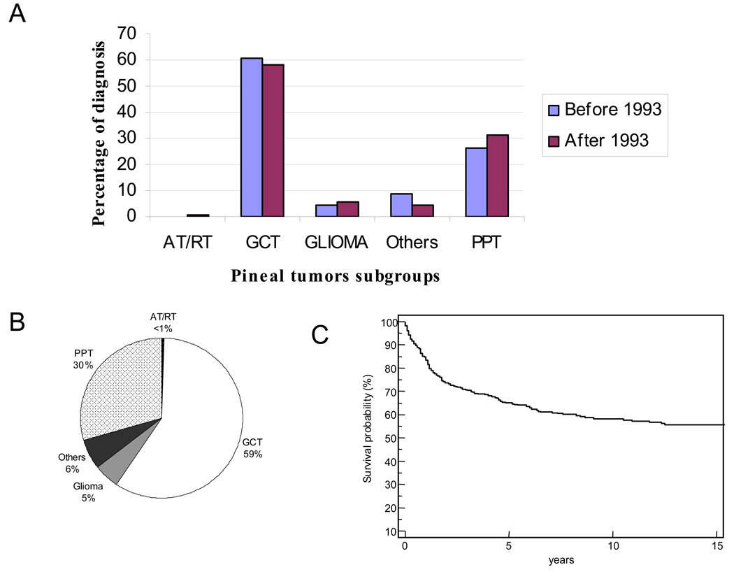
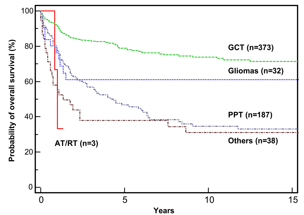
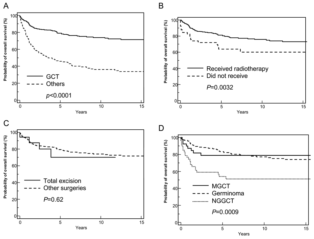
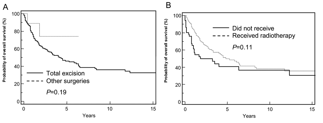
Pineal gland tumors are rare and account for less than 1% of all primary brain tumor diagnoses. They are more commonly seen in pediatric patients than in adults. We analyzed the available SEER data on pineal gland tumors that were diagnosed during the period 1973-2005. The cohort was subdivided into groups on the basis of tumor histology: germ cell tumors, pineal parenchymal tumors, gliomas, and other pineal tumors. Analyses of incidence, survival, factors influencing survival, and treatment modalities are provided. Among the 633 patients with pineal tumors, male sex was predominant, i.e., sex ratio was 3:1 for the whole group and 11.8:1 for those with germ cell tumors. The 5-year overall survival (OS) for the cohort was 65% +/- 2.1%. Those with germ cell tumors experienced the best survival (OS = 78.9% +/- 2.3%), followed by those with gliomas (OS = 61% +/- 9.3%), and those with pineal parenchymal tumors (OS = 47.2% +/- 4.2%). Non-germ cell tumors, absence of radiotherapy from treatment regimen, and diagnosis before 1993 were the only factors associated with a negative impact on survival. The extent of surgical tumor resection did not affect survival in any histologic subgroup. We conclude that, although pineal tumors are histologically diverse, they share some similarities due to their unique location. An aggressive surgical approach should be considered with caution in this region. Further studies on different pineal tumors subtypes are needed.
Figures
References
-
- Cbtrus. Statistical Report: Primary Brain Tumors In The United States -Pbtcbtrotus. 2005. - PubMed
-
- Brat DJ PJ, Kleinschmidt-Demasters BK, Yachnis AT, Montine TJ, Boyer PJ, Powell SZ, Prayson RA, Mclendon RE. Neuropathology Committee, College Of American Pathologists.: Surgical Neuropathology Update. A Review Of Changes Introduced By The Who Classification Of Tumours Of The Central Nervous System. Arch Pathol Lab Med. (4th Edition.) 2008:993–1007. - PubMed
-
- Kersh CR CW, Eisert DR, Spaulding CA, Hahn SS, Jenrette JM, 3rd, Marks RD., Jr Primary Central Nervous System Germ Cell Tumors. Effect Of Histologic Confirmation On Radiotherapy. Cancer. 1988;61:2148–2142. 2152. - PubMed
-
- Maria E.Echeverria Jf, Goldman Stewart. Pediatric Central Nervous System Germ Cell Tumors: A Review. The Oncologist. 2008;13:690–699. - PubMed
-
- Roger J. Packer TM, Vezina Gilbert. Central Nervous System Tumors. Pediatr Clin N Am. 2008;55:121–145. - PubMed
Publication types
MeSH terms
Grants and funding
LinkOut - more resources
Full Text Sources
Medical
