Review
. 2008;39(1-4):149-58.
doi: 10.1007/s12029-009-9058-y. Epub 2009 Apr 17.

The tumor suppressor function of human sulfatase 1 (SULF1) in carcinogenesis

Affiliations
Review

The tumor suppressor function of human sulfatase 1 (SULF1) in carcinogenesis

Jin-Ping Lai et al. J Gastrointest Cancer. 2008.

Abstract

Introduction: Human sulfatase 1 (SULF1) was recently identified and shown to desulfate cellular heparan sulfate proteoglycans (HSPGs). Since sulfated HSPGs serve as co-receptors for many growth factors and cytokines, SULF1 was predicted to modulate growth factor and cytokine signaling.

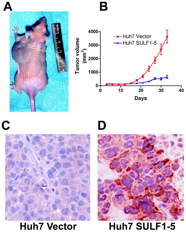
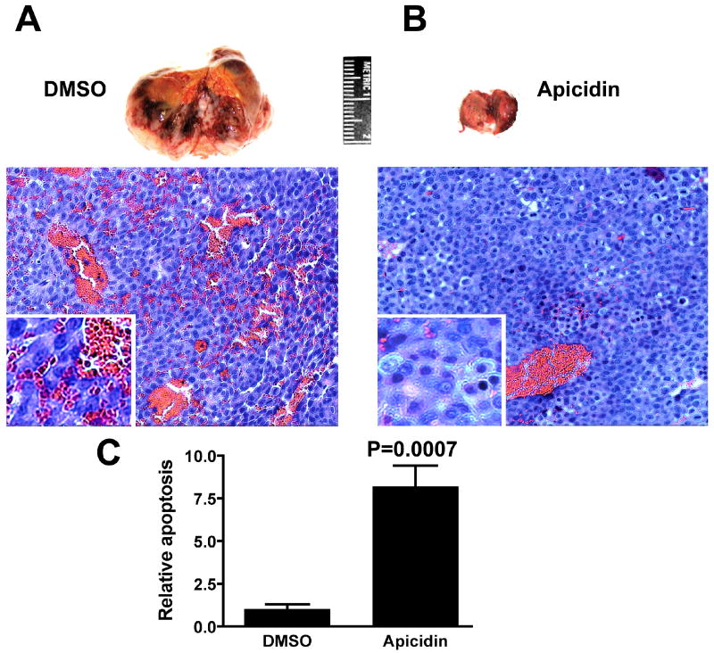
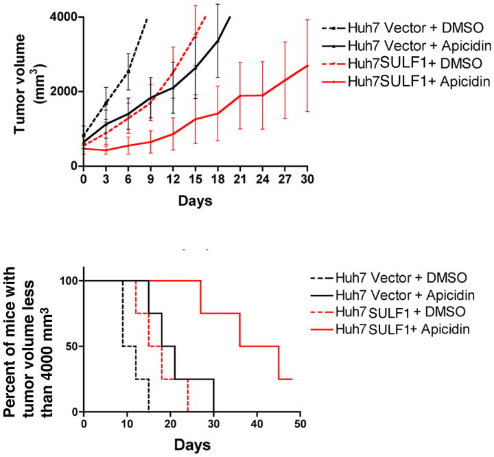
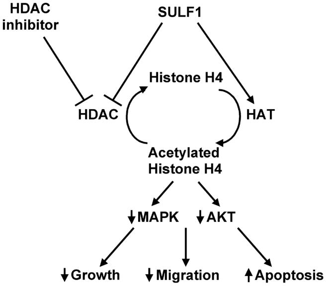
Discussion: The role of SULF1 in growth factor signaling and its effects on human tumorigenesis are under active investigation. Initial results show that SULF1 inhibits the co-receptor function of HSPGs in multiple receptor tyrosine kinase signaling pathways, particularly by the heparin binding growth factors--fibroblast growth factor 2, vascular endothelial growth factor, hepatocyte growth factor, PDGF, and heparin-binding epidermal growth factor (HB-EGF). SULF1 is downregulated in the majority of cancer cell lines examined, and forced expression of SULF1 decreases cell proliferation, migration, and invasion. SULF1 also promotes drug-induced apoptosis of cancer cells in vitro and inhibits tumorigenesis and angiogenesis in vivo.

Conclusion: Strategies targeting SULF1 or the interaction between SULF1 and the related sulfatase 2 will potentially be important in developing novel cancer therapies.


Figures

Figure 1
Figure 1
Forced expression of SULF1 significantly delays growth of HCC xenografts in nude mice. A: Two million Huh7 cells stably transfected with empty plasmid vector (injected into the left side of the mouse in) or with a plasmid expressing the full length SULF1 (injected into the right side of the mouse) were inoculated subcutaneously into the flanks of nude mice and the resulting xenografts photographed on the 30th day after inoculation. B: The graph shows the results from xenografts implanted into 10 nude mice; there was profound inhibition of growth of Huh7 xenografts expressing SULF1 (P<0.001). C & D: Immunohistochemistry was performed to confirm human SULF1 protein expression in the xenografts from cell lines stably-transfected with empty vector (C) and SULF1 (D) using a rabbit polyclonal antibody against SULF1.
Figure 2
Figure 2
Apicidin inhibits growth and induces apoptosis in Huh7 xenografts in nude mice. A and B: Five million Huh7 cells were inoculated subcutaneously into the right flanks of nude mice. When tumor sizes reached 400-800 mm3, the mice were randomly grouped and treated by intraperitoneal injection of either 1% DMSO or apicidin at 2.5 mg/kg daily for the first week and every other day for the second week. Nude mice bearing xenografts were then sacrificed. Tissue from the xenografts was fixed with formalin and paraffin embedded. After H & E staining, the percent apoptosis in 500 nuclei from 6 randomly selected areas of each slide was counted. The relative rate of apoptosis in DMSO vs. apicidin treated mice was graphed. C: Apicidin significantly induced apoptosis in Huh7 xenografts (P=0.0007).
Figure 3
Figure 3
SULF1 enhances the efficacy of apicidin in growth inhibition of HCC xenografts. Huh7 xenografts with or without SULF1 expression were generated. When the volume of xenografts reached 400 to 800 mm3, the nude mice were grouped randomly and treated with 100 μl 10% DMSO for controls and 100 μl of apicidin dissolved in 10% DMSO at a total dose of 2.5 mg/kg body weight. The mice were treated IP daily for one week, every other day for one week and every three days for two weeks. Tumor size was measured with calipers every three days and the mice were sacrificed when their tumor volume reached 4000 mm3. The combination of SULF1 and apicidin inhibits tumor growth in mice bearing Huh7 xenografts.
Figure 4
Figure 4
Working Model for the effect of SULF1 and HDAC inhibitors on histone acetylation and downstream pathways. Forced expression of SULF1 disrupts the balance between HDACs and HATs by inhibiting HDACs and/or by up-regulating HAT activity. This leads to an increase in the acetylation of histone H4 and to subsequent down-regulation of AKT and MAPK kinase pathways. Therefore, SULF1, potentiates the effects of HDAC inhibitors both in vitro and in vivo.
Figure 5
Figure 5
Additive effect of apicidin and doxorubicin (DOX) in SULF1-expressing HCC cells in vivo. Huh7 xenografts with or without SULF1 expression were generated. When the volume of xenografts reached 400 to 800 mm3, the nude mice were grouped randomly and treated with 100 μl 10% DMSO for controls, 100 μl of apicidin dissolved in 10% DMSO at a dose of 2.5 mg/kg body weight, 100 μl of DOX at a dose of 3 mg/kg body weight, or the combination of apicidin with DOX. The mice were treated IP with DMSO or apicidin daily for one week, every other day for one week and every three days for two weeks, and IV with DOX via tail vein every 6 days for four times. Tumor size was measured with calipers every three days and the mice were sacrificed when their tumor volume reached 4000 mm3. The experiments were repeated and the mice were sacrificed at the end of the fourth week's treatment. Compared to the DMSO control, DOX inhibited tumor growth of Huh7 vector xenografts (P<0.05), but these effects were not statistically further enhanced by combination of the DOX with apicidin (A and C) . Compared to the Huh7 vector xenografts, Huh7 SULF1 expressing xenografts showed more profound inhibition of tumor growth after treatment with DOX (P<0.05). Further, in SULF1-expressing Huh7 xenografts the combination of apicidin with DOX showed significantly enhanced anti-tumor effects as compared with the effect of either apicidin or DOX alone (P<0.05) (B and D).

References

    1. Diez-Roux G, Ballabio A. Sulfatases and human disease. Annu Rev Genomics Hum Genet. 2005;6:355–379. - PubMed
    1. Shridhar V, Sen A, Chien J, Staub J, Avula R, Kovats S, Lee J, Lillie J, Smith DI. Identification of underexpressed genes in early- and late-stage primary ovarian tumors by suppression subtraction hybridization. Cancer research. 2002;62(1):262–270. - PubMed
    1. Ai X, Do AT, Kusche-Gullberg M, Lindahl U, Lu K, Emerson CP., Jr Substrate specificity and domain functions of extracellular heparan sulfate 6-O-endosulfatases, QSulf1 and QSulf2. The Journal of biological chemistry. 2006;281(8):4969–4976. - PubMed
    1. Dhoot GK, Gustafsson MK, Ai X, Sun W, Standiford DM, Emerson CP., Jr Regulation of Wnt signaling and embryo patterning by an extracellular sulfatase. Science. 2001;293(5535):1663–1666. - PubMed
    1. Lai J, Chien J, Staub J, Avula R, Greene EL, Matthews TA, Smith DI, Kaufmann SH, Roberts LR, Shridhar V. Loss of HSulf-1 up-regulates heparin-binding growth factor signaling in cancer. The Journal of biological chemistry. 2003;278(25):23107–23117. - PubMed

Publication types

MeSH terms

Substances