. 2009;61(3):322-31.
doi: 10.1080/01635580802521338.

Sodium selenite increases the activity of the tumor suppressor protein, PTEN, in DU-145 prostate cancer cells

Affiliations

Sodium selenite increases the activity of the tumor suppressor protein, PTEN, in DU-145 prostate cancer cells

Margareta Berggren et al. Nutr Cancer. 2009.

Abstract

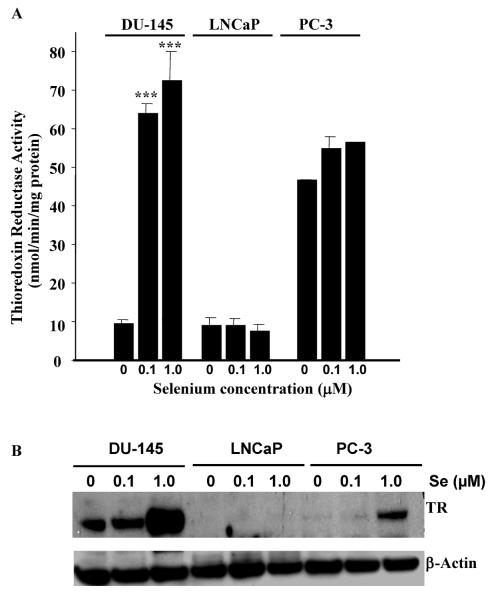
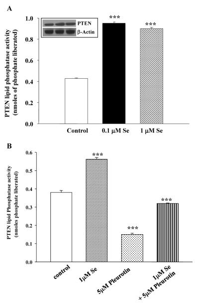
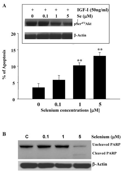
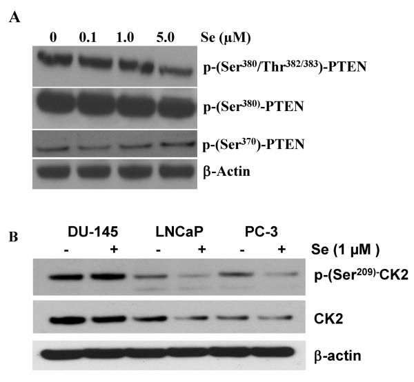
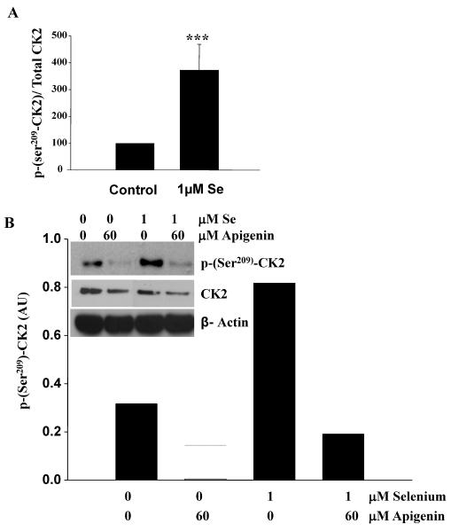
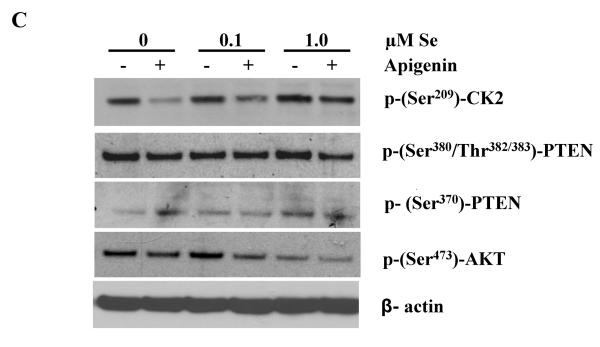
Epidemiological and clinical data suggest that selenium may prevent prostate cancer; however, the cellular effects of selenium in malignant prostate cells are not well understood. We previously reported that the activity of the tumor suppressor PTEN is modulated by thioredoxin (Trx) in a RedOx-dependent manner. In this study, we demonstrated that the activity of Trx reductase (TR) is increased by sevenfold in the human prostate cancer cell line, DU-145, after 5 days of sodium selenite (Se) treatment. The treatment of DU-145 cells with increasing concentrations of Se induced an increase in PTEN lipid phosphatase activity by twofold, which correlated with a decrease in phospho-ser(473)-Akt, and an increase in phospho-Ser(370)-PTEN levels. Se also increased casein kinase-2 (CK2) activity; and the use of apigenin, an inhibitor of CK2, revealed that the regulation of the tumor suppressor PTEN by Se may be achieved via both the Trx-TR system and the RedOx control of the kinase involved in the regulation of PTEN activity.


Figures

Figure 1
Figure 1. Thioredoxin reductase activity is increased by sodium selenite in DU-145 prostate cancer cells
Prostate cancer cells (DU-145, PC-3 and LNCaP) were treated for 5 days with 0.1 or 1.0μM sodium selenite. TR activity was measured as described in the Materials and Methods Section (Panel A). TR was increased significantly in a dose dependent manner by sodium selenite in DU-145 cells. Bars represent means ± SE from two independent experiments performed in triplicate (n=6). Significance was achieved with ** for p<0.01 and *** for p<0.001. Panel B represents the expression of TR in DU-145, PC-3 and LNCaP prostate cancer cells following 0.1 and 1.0 μM sodium selenite treatment. Note the large increase in TR protein expression in DU-145 cells as compared to LNCaP and PC-3 upon sodium 1.0 μM selenite treatment.
Figure 2
Figure 2. Effect of sodium selenite on glutathione peroxidase activity
DU-145, PC-3 and LNCaP prostate cancer cells were treated with various concentrations of sodium selenite for 5 days at the indicated concentrations. The graph represents the activity of glutathione peroxidase as measured by the amount of reduced NADPH produced/min/mg of total protein/sample. Note that the level of glutathione peroxidase activity remains low in DU-145 and LNCaP cells as compared to the level in PC-3 cells.
Figure 3
Figure 3. Effect of sodium selenite on Trx and GSH bioreduction capacity
DU-145 cells were treated with various concentrations of sodium selenite for 5 days at the indicated concentrations. Panel A, DU-145 cells were treated with sodium selenite for 5 days and BSO at 1mM for 1 hour prior to the bioreduction assay. Cells were incubated with buffer containing 5mM glucose plus vehicle, or 5mM oxidized lipoate. The rate of Trx bioreduction was determined using 6 measurements over a 1hr period in duplicate treatment samples. Note that Trx's bioreduction potential increased significantly upon sodium selenite treatment. Bars represent means ± SE from three independent experiments performed in triplicate. Significance was achieved with *** represents significance at p< 0.001, when compared to respective vehicles or # represents significance at p< 0.01, when compared to lipoate reduction in control. Panel B, DU-145 cells were treated with sodium selenite for 5 days then cells were incubated with buffer containing 5mM glucose plus vehicle or 5mM HEDS. Note that there was no affect of selenite on GSH bioreduction. Bars represent means ± SE from three independent experiments performed in duplicate.
Figure 4
Figure 4. PTEN lipid phosphastase is increased by sodium selenite via the Trx-TR system
Panel A represents PTEN lipid phosphatase activity as measured by the release of nmoles of phosphate from the substrate PtdIns-3,4,5-P3 as described in the Materials and Methods section. Note that increasing concentrations of sodium selenite stimulates PTEN lipid phosphatase activity in DU-145 cells. Inset shows the total PTEN protein expression in the corresponding samples as compared to internal control, β-actin. Panel B represents PTEN activity following 1μM sodium selenite, 5μM pleurotin (an inhibitor of TR) or the combination treatment. The combination treatment restores PTEN activity back to basal activity as compared to that in control cells. Bars represent means ± SE from three independent experiments performed in triplicate. Significance was achieved with *** for p<0.001.
Figure 5
Figure 5. Sodium selenite inhibits Akt activation, induces apoptosis and PARP cleavage in DU-145 prostate cancer cells
The insert of Panel A represents a typical western blot for the inhibition of Akt phosphorylation by increasing concentrations of sodium selenite in DU-145 cells. Briefly, cells were lysed and phosphorylated Akt was detected using specific anti-phospho-Ser473-Akt. Akt phosphorylation was decreased in the presence of increasing concentration of sodium selenite in DU-145 cells, correlating with the increase in PTEN lipid phosphastase activity. The graph in Panel A represents the induction of apoptosis in DU-145 cells by selenite as measured by the morphological assay described in the Materials and Methods section. Bars represent means ± SE from two independent experiments performed in triplicate. Significance was achieved with ** for p<0.01. Panel B represents a typical western blot for the cleavage of PARP by increasing concentrations of sodium selenite in DU-145 cells. Actin was used a loading control. Note the lower band appearing in cells treated with 5 μM selenite, which corresponds to cleaved PARP and induction of apoptosis (Panel A).
Figure 6
Figure 6. Sodium selenite modulated PTEN phosphorylation in DU-145 cells
DU-145 cells were treated with increasing concentrations of sodium selenite for 5 days. Cells were lysed and samples were probed for PTEN phosphorylation using specific phospho-antibodies. Panel A represents a typical Western blot for PTEN phosphorylation on residues Ser380/Thr382/383, PTEN phosphorylation on Ser380 and on Ser370 as compared to loading control with β-actin. Note the decrease in PTEN phosphorylation with 5μM of Sodium selenite on Ser380/Thr382/383 and a slight increase in PTEN phosphorylation on Ser370. Panel B represents the expression and the phosphorylation status of CK2 involved in the phosphorylation of PTEN following increasing concentrations of sodium selenite in DU-145, LNCaP and PC-3 prostate cancer cells. Cells were treated with 1μM of Se. Note the high levels of CK2 and phospho-(Ser209)-CK2 in DU-145 cells as compared to the other cell lines.
Figure 7
Figure 7. Sodium selenite increases the activity of CK2 in DU-145 cells
DU-145 cells were treated with different concentrations of sodium selenite for 5 days. Cells were lysed and samples were probed for phospho-Ser209CK2 and total CK2 using specific phospho-and total antibodies. Panel A represents the quantification of five separate experiments. Note the significant increase in the ratio phospho-Ser209CK2/Total CK2 following Se treatment in DU-145 cells. Bars represent means ± SE from five independent experiments performed in triplicate. Significance was achieved with *** for p<0.001. The inset in Panel B represents a typical western blot, where phospho-Ser209-CK2, total CK2 and β-actin were probed following treatment of the cells with Se with or without 60μM of apigenin. The graph represents the quantification of the typical western blot shown in the inset. Note that apigenin counteracts the effects of Se on CK2 phosphorylation. Panel C represents a typical western blot from DU-145 cells that were pre-treated with increasing concentrations of Se and then incubated with apigenin or vehicle control. Specific anti-phospho-Ser209CK2, phospho-Ser473Akt, phospho-Ser370PTEN and phospho-Ser380/Thr382/383PTEN antibodies were used to probe the membrane. Note that Se induces CK2 and PTEN phosphorylation (Ser380/Thr382/383) and decrease Akt phosphorylation. Se can also partially rescue the effects of apigenin on CK2.

Similar articles

Cited by

References

    1. Linder MC. Nutrition and metabolism trace elements. In: Linder MC, editor. Nutritional biochemistry and metabolism with clinical applications. Elsevier; New York: 1990.
    1. Jiang C, Wang Z, Ganther H, Lu J. Distinct effects of methylseleninic acid versus selenite on apoptosis, cell cycle, and protein kinase pathways in DU145 human prostate cancer cells. Mol Cancer Ther. 2002;1:1059–1066. - PubMed
    1. Wang Z, Jiang C, Lu J. Induction of caspase-mediated apoptosis and cell-cycle G1 arrest by selenium metabolite methylselenol. Mol Carcinog. 2002;34:113–12. - PubMed
    1. Wu Y, Zu K, Warren MA, Wallace PK, Ip C. Delineating the mechanism by which selenium deactivates Akt in prostate cancer cells. Mol Cancer Ther. 2006;5:246–252. - PubMed
    1. Lee YC, Tang YC, Chen YH, Wong CM, Tsou AP. Selenite-induced survival of HuH7 hepatoma cells involves activation of focal adhesion kinase-phosphatidylinositol 3-kinase-Akt pathway and Rac1. J Biol Chem. 2003;278:39615–39624. - PubMed

MeSH terms