Comparative Study
. 2009 Sep;220(3):586-92.
doi: 10.1002/jcp.21735.

Survival responses of human embryonic stem cells to DNA damage

Affiliations
Comparative Study

Survival responses of human embryonic stem cells to DNA damage

Tera M Filion et al. J Cell Physiol. 2009 Sep.

Abstract

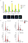
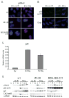
Pluripotent human embryonic stem (hES) cells require mechanisms to maintain genomic integrity in response to DNA damage that could compromise competency for lineage-commitment, development, and tissue-renewal. The mechanisms that protect the genome in rapidly proliferating hES cells are minimally understood. Human ES cells have an abbreviated cell cycle with a very brief G1 period suggesting that mechanisms mediating responsiveness to DNA damage may deviate from those in somatic cells. Here, we investigated how hES cells react to DNA damage induced by ionizing radiation (IR) and whether genomic insult evokes DNA repair pathways and/or cell death. We find that hES cells respond to DNA damage by rapidly inducing Caspase-3 and -8, phospho-H2AX foci, phosphorylation of p53 on Ser15 and p21 mRNA levels, as well as concomitant cell cycle arrest in G2 based on Ki67 staining and FACS analysis. Unlike normal somatic cells, hES cells and cancer cells robustly express the anti-apoptotic protein Survivin, consistent with the immortal growth phenotype. SiRNA depletion of Survivin diminishes hES survival post-irradiation. Thus, our findings provide insight into pathways and processes that are activated in human embryonic stem cells upon DNA insult to support development and tissue regeneration.


Figures
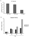
Fig. 1
Fig. 1
Decreased cell survival of human ES cells upon γ-irradiation. A. Survival of H1 cells was determined by assessing the number of adherent live cells (as determined by trypan blue staining) per well of a 6-well plate before and at 7 h after IR with 5 Gy. Values represent the average of three determinations within a representative experiment and the error bars are based on the standard error. B. Induction of apoptosis was determined by monitoring the activities of Caspase-3 and Caspase-8 by enzymatic assays at 2, 3 and 4 h after IR. Values represent the average of sample duplicates. Standard error was determined, but this error is minimal and not readily visible in the graph).
Fig. 2
Fig. 2
γ-Irradiation induces a G2 phase related cell cycle arrest in human ES cells. A. Immuno-fluorescence microscopy was used to examine Ki67 staining patterns (n=200; bar graph in upper portion), which reflect different stages of the cell cycle (lower portion), as a time-course after γ-irradiation (5 Gy). Values represent the average of three independent determinations of 200 cells per sample within a representative experiment and the error bars are based on the standard error. B. Fluorescence activated cell sorting was used to assess the distribution of hES cells in different cell cycle stages following γ-irradiation. We note that the fraction of cells in G1 may be over-estimated due to the presence of inactivated mouse embryonic fibroblasts that are arrested in G1.
Fig. 3
Fig. 3
Activation of the ATM pathway activation by IR in hES cells. A. Immuno-fluorescence microscopy was used to examine the in situ appearance of γH2A.X foci, which reflect phosphorylation of H2A.X by ATM at sites of double-stranded DNA breaks after IR. B. Same as A, but assessing phosphorylation of p53 at Ser-15 by in situ immunofluorescence microscopy after IR. C. Quantitative real-time PCR analysis was used to show that in situ activation of the ATM pathway after IR results in the expected p53 mediated induction of p21 mRNA levels at 2 and 7 h after radiation with 5 Gy. Error bars represent standard deviation of sample duplicates. D. Western blot analysis was used to show activation of components of the ATM pathway in human H1 embryonic stem cells, normal diploid WI-38 fibroblasts and MDA-MB-231 breast cancer cells upon γ-irradiation. Blots were prepared with antibodies probing ATM dependent phosphorylation of H2A.X (γH2A.X) and Ser-15 phosphorylation of p53, as well as the levels of the CDK inhibitor p21 and Chk2, using α-tubulin as an internal control for protein loading.
Fig. 4
Fig. 4
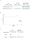
Expression and function of survivin upon γ-irradiation in human ES cells. A. Western blot analysis was used to monitor survivin protein levels in human H1 embryonic stem cells, normal diploid WI-38 fibroblasts and MDA-MB-231 breast cancer cells upon γ-irradiation (5 Gy) at the indicated time-points. Levels of α-tubulin were determined as an internal control for protein loading. B. Effect of siRNA-mediated depletion of survivin during the DNA damage response in human ES cells. Cells were transfected with either control or survivin siRNA for 48 hours and then exposed to 5Gy of γ-irradiation for 7 min. siRNA treatment is effective as revealed by reduced survivin levels throughout a 96 h time course after IR (lower portions of Panels B and C). Cell survival was measured by MTS assay as function of time after treatment (graphs in upper portions of Panels B and C).

References

    1. Abdul-Ghani M, Megeney LA. Rehabilitation of a contract killer: caspase-3 directs stem cell differentiation. Cell Stem Cell. 2008;2:515–516. - PubMed
    1. Aladjem MI, Spike BT, Rodewald LW, Hope TJ, Klemm M, Jaenisch R, Wahl GM. ES cells do not activate p53-dependent stress responses and undergo p53-independent apoptosis in response to DNA damage. Curr Biol. 1998;8:145–155. - PubMed
    1. Altieri DC. Survivin, versatile modulation of cell division and apoptosis in cancer. Oncogene. 2003;22:8581–8589. - PubMed
    1. Altieri DC. The case for survivin as a regulator of microtubule dynamics and cell-death decisions. Curr Opin Cell Biol. 2006;18:609–615. - PubMed
    1. Altieri DC. New wirings in the survivin networks. Oncogene. 2008a;27:6276–6284. - PMC - PubMed

Publication types

MeSH terms

Substances

LinkOut - more resources