C-peptide of preproinsulin-like peptide 7: localization in the rat brain and activity in vitro
- PMID: 19373968
- PMCID: PMC2775513
- DOI: 10.1016/j.neuroscience.2009.01.031
C-peptide of preproinsulin-like peptide 7: localization in the rat brain and activity in vitro
Abstract
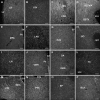
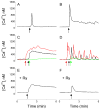
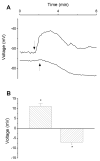
With the use of a rabbit polyclonal antiserum against a conserved region (54-118) of C-peptide of human preproinsulin-like peptide 7, referred to herein as C-INSL7, neurons expressing C-INSL7-immunoreactivity (irC-INSL7) were detected in the pontine nucleus incertus, the lateral or ventrolateral periaqueductal gray, dorsal raphe nuclei and dorsal substantia nigra. Immunoreactive fibers were present in numerous forebrain areas, with a high density in the septum, hypothalamus and thalamus. Pre-absorption of C-INSL7 antiserum with the peptide C-INSL7 (1 microg/ml), but not the insulin-like peptide 7 (INSL7; 1 microg/ml), also known as relaxin 3, abolished the immunoreactivity. Optical imaging with a voltage-sensitive dye bis-[1,3-dibutylbarbituric acid] trimethineoxonol (DiSBAC4(3)) showed that C-INSL7 (100 nM) depolarized or hyperpolarized a small population of cultured rat hypothalamic neurons studied. Ratiometric imaging studies with calcium-sensitive dye fura-2 showed that C-INSL7 (10-1000 nM) produced a dose-dependent increase in cytosolic calcium concentrations [Ca2+]i in cultured hypothalamic neurons with two distinct patterns: (1) a sustained elevation lasting for minutes; and (2) a fast, transitory rise followed by oscillations. In a Ca2+-free Hanks' solution, C-INSL7 again elicited two types of calcium transients: (1) a fast, transitory increase not followed by a plateau phase, and (2) a transitory rise followed by oscillations. INSL7 (100 nM) elicited a depolarization or hyperpolarization in a small population of hypothalamic neurons, and an increase of [Ca2+]i with two patterns that were dissimilar from that of C-INSL7. [125I]C-INSL7 bindings to rat brain membranes were inhibited by C-INSL7 in a dose-dependent manner; the Kd and Bmax. values were 17.7 +/- 8.2 nM and 45.4 +/- 20.5 fmol/mg protein. INSL7 did not inhibit [125I]C-INSL7 binding to rat brain membranes, indicating that C-INSL7 and INSL7 bind to distinct binding sites. Collectively, our result raises the possibility that C-INSL7 acts as a signaling molecule independent from INSL7 in the rat CNS.
Figures


Similar articles
-
Neuronostatin is co-expressed with somatostatin and mobilizes calcium in cultured rat hypothalamic neurons.Neuroscience. 2010 Mar 17;166(2):455-63. doi: 10.1016/j.neuroscience.2009.12.059. Epub 2010 Jan 4. Neuroscience. 2010. PMID: 20056135 Free PMC article.
-
Relaxin-3 in GABA projection neurons of nucleus incertus suggests widespread influence on forebrain circuits via G-protein-coupled receptor-135 in the rat.Neuroscience. 2007 Jan 5;144(1):165-90. doi: 10.1016/j.neuroscience.2006.08.072. Epub 2006 Oct 30. Neuroscience. 2007. PMID: 17071007
-
Identification of relaxin-3/INSL7 as an endogenous ligand for the orphan G-protein-coupled receptor GPCR135.J Biol Chem. 2003 Dec 12;278(50):50754-64. doi: 10.1074/jbc.M308995200. Epub 2003 Sep 30. J Biol Chem. 2003. PMID: 14522968
-
KiSS-1 expression and metastin-like immunoreactivity in the rat brain.J Comp Neurol. 2005 Jan 17;481(3):314-29. doi: 10.1002/cne.20350. J Comp Neurol. 2005. PMID: 15593369
-
Excitatory effects of human immunodeficiency virus 1 Tat on cultured rat cerebral cortical neurons.Neuroscience. 2008 Feb 6;151(3):701-10. doi: 10.1016/j.neuroscience.2007.11.031. Epub 2007 Dec 5. Neuroscience. 2008. PMID: 18164555 Free PMC article.
Cited by
-
Direct evidence of intracrine angiotensin II signaling in neurons.Am J Physiol Cell Physiol. 2014 Apr 15;306(8):C736-44. doi: 10.1152/ajpcell.00131.2013. Epub 2014 Jan 8. Am J Physiol Cell Physiol. 2014. PMID: 24401846 Free PMC article.
-
Hyperinsulinemia in Obesity, Inflammation, and Cancer.Diabetes Metab J. 2021 May;45(3):285-311. doi: 10.4093/dmj.2020.0250. Epub 2021 Mar 29. Diabetes Metab J. 2021. PMID: 33775061 Free PMC article. Review.
References
-
- Adham IM, Burkhardt E, Benahmed M, Engel W. Cloning of a cDNA for a novel insulin-like peptide of the testicular Leydig cells. J Biol Chem. 1993;268:26668–26672. - PubMed
-
- Bathgate RA, Samuel CS, Burazin TC, Layfield S, Claasz AA, Reytomas IG, Dawson NF, Zhao C, Bond C, Summers RJ, Parry LJ, Wade JD, Tregear GW. Human relaxin gene 3 (H3) and the equivalent mouse relaxin (M3) gene. Novel members of the relaxin peptide family. J Biol Chem. 2002;277:1148–1157. - PubMed
-
- Bell GI, Merryweather JP, Sanchez-Pescador R, Stempien MM, Priestley L, Scott J, Rall LB. Sequence of a cDNA clone encoding human preproinsulin-like growth factor II. Nature. 1984;310:775–777. - PubMed
-
- Brailoiu E, Churamani D, Pandey V, Brailoiu GC, Tuluc F, Patel S, Dun NJ. Messenger-specific role for nicotinic acid adenine dinucleotide phosphate in neuronal differentiation. J Biol Chem. 2006;281:15923–15928. - PubMed
Publication types
MeSH terms
Substances
Grants and funding
LinkOut - more resources
Full Text Sources
Miscellaneous

