. 2009 Nov;15(11):3221-30.
doi: 10.1089/ten.TEA.2008.0545.

Photocrosslinking of gelatin macromers to synthesize porous hydrogels that promote valvular interstitial cell function

Affiliations

Photocrosslinking of gelatin macromers to synthesize porous hydrogels that promote valvular interstitial cell function

Julie A Benton et al. Tissue Eng Part A. 2009 Nov.

Abstract

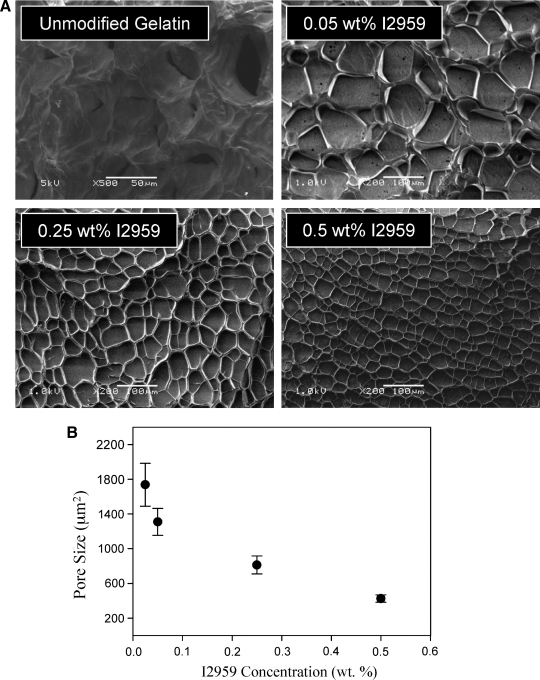
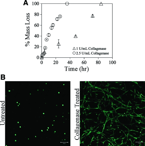
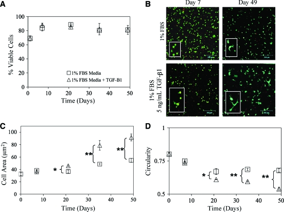
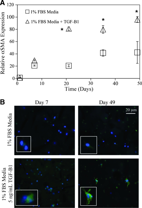
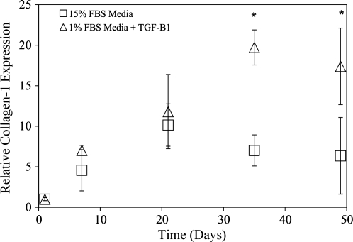
The development of novel three-dimensional cell culture platforms for the culture of aortic valvular interstitial cells (VICs) has been fraught with many challenges. Although the most tunable, purely synthetic systems have not been successful at promoting cell survivability or function. On the other hand, entirely natural materials lack mechanical integrity. Here we explore a novel hybrid system consisting of gelatin macromers synthetically modified with methacrylate functionalities allowing for photoencapsulation of cells. Scanning electron microscopy observations show a microporous structure induced during polymerization within the hydrogel. This porous structure was tunable with polymerization rate and did not appear to have interconnected pores. Treatment with collagenase caused bulk erosion indicating enzymatic degradation controls the matrix remodeling. VICs, an important cell line for heart valve tissue engineering, were photoencapsulated and examined for cell-directed migration and differentiation. VICs were able to achieve their native morphology within 2 weeks of culture. The addition of the pro-fibrotic growth factor, transforming growth factor-beta1, accelerated this process and also was capable of inducing enhanced alpha-smooth muscle actin and collagen-1 expression, indicating a differentiation from quiescent fibroblasts to active myofibroblasts as demonstrated by quantitative real-time polymerase chain reaction and immunohistochemistry. Although these studies were limited to VICs, this novel hydrogel system may also be useful for studying other fibroblastic cell types.


Figures

FIG. 1.
FIG. 1.
(A) Scanning electron microscopic images of 10 wt% lyophilized methacrylated gelatin macromer (GelMA) hydrogel samples with varying photoinitiator concentration and natural gelatin gel. (B) Quantification of GelMA hydrogel porosity from scanning electron microscopic images as a function of initiator concentration. Porosity was determined using the projected area of the pores as determined with NIH ImageJ software. The largest pores were achieved with the least amount of photoinitiator, while the smallest pores were formed with most amount of photoinitiator. Results were calculated with three or more electron micrographs per sample.
FIG. 2.
FIG. 2.
(A) GelMA 10 wt% hydrogels of uniform size were exposed to 2.5 U/mL (○) or 1 U/mL (▵) exogenous collagenase until complete degradation. The higher concentration of collagenase increased the rate of degradation as expected. (B) Valvular interstitial cells (VICs) were encapsulated in 10 wt% GelMA hydrogels and incubated with or without 1 U/mL of exogenous collagenase for 4 h. Collagenase was then removed, and the cells were cultured for an additional 2 days. GelMA-VIC hydrogels were then stained with Live/Dead® and imaged with a confocal microscope. No dead cells were observed. Scale bar = 100 μm. VICs had more dramatic spread morphology in treated hydrogels compared with untreated hydrogels. Color images available online at www.liebertonline.com/ten.
FIG. 3.
FIG. 3.
(A) VIC viability was measured with Live/Dead staining over a time course of 7 weeks. Constructs were treated with (▵) or without (□) 5 ng/mL transforming growth factor-β1 (TGF-β1) in 1% fetal bovine serum (FBS) medium. Initially, the viability of encapsulated VICs dropped, and then recovered after 1 week in culture. Growth factor treatment had no apparent effect on overall cell viability. (B) Representative Live/Dead projected confocal stacks of VICs encapsulated within GelMA hydrogel networks treated with TGF-β1 as indicated. Greater cell spreading was observed in cells treated with TGF-β1. Scale bar = 200 μm. (C) To quantify differences in cell spreading, average cell area was measured from Live/Dead images with NIH ImageJ software. Significant differences in cell spreading were observed from day 21 onward with TGF-β1 treatment. (D) To confirm differences in cell spreading, circularity was also measured from Live/Dead images with NIH ImageJ software. Significant differences in circularity were observed from day 21 onward with TGF-β1 treatment. n = 3, *p < 0.01, **p < 0.001. Color images available online at www.liebertonline.com/ten.
FIG. 4.
FIG. 4.
(A) Quantitative real-time polymerase chain reaction analysis of α smooth muscle actin (αSMA) messenger RNA (mRNA) expression treated with (▵) or without (□) 5 ng/mL TGF-β1 in 1% FBS medium. Expression indicated is relative to the day 1 time point and glyceraldehyde 3-phophate dehydrogenase housekeeping gene expression. αSMA mRNA was elevated by twofold with TGF-β1 treatment. (B) Immunohistochemical staining of αSMA protein expression at days 7 and 49 in 30 μm cryosections. Positive αSMA staining is shown in green; 4′,6′-diamidino-2-phenylindole (DAPI) counterstaining for cell nuclei is depicted in blue. Cells treated with TGF-β1 had greater staining for αSMA with distinct stress fiber formation, indicating myofibroblastic differentiation. A blow-up of stress fiber positive cells is also shown as an inset. *p ≤ 0.01 over the untreated VIC-GelMA constructs, n = 4 for all samples.
FIG. 5.
FIG. 5.
Quantitative real-time polymerase chain reaction analysis of collagen-1 mRNA expression treated with (▵) or without (□) 5 ng/mL TGF-β1 in 1% FBS medium. Expression indicated is relative to the day 1 time point and glyceraldehyde 3-phophate dehydrogenase housekeeping gene expression. Collagen-1 mRNA was elevated by 1.5-fold with TGF-β1 treatment. *p ≤ 0.01 over the untreated VIC-GelMA constructs, n = 4 for all samples.

References

    1. Abbott A. Cell culture: biology's new dimension. Nature. 2003;424:870. - PubMed
    1. Raeber G.P. Lutolf M.P. Hubbell J.A. Molecularly engineered PEG hydrogels: a novel model system for proteolytically mediated cell migration. Biophys J. 2005;89:1374. - PMC - PubMed
    1. Salinas C.N. Anseth K.S. The enhancement of chondrogenic differentiation of human mesenchymal stem cells by enzymatically regulated RGD functionalities. Biomaterials. 2008;29:2370. - PMC - PubMed
    1. Nuttelman C.R. Tripodi M.C. Anseth K.S. In vitro osteogenic differentiation of human mesenchymal stem cells photoencapsulated in PEG hydrogels. J Biomed Mater Res A. 2004;68A:773. - PubMed
    1. Nuttelman C.R. Tripodi M.C. Anseth K.S. Synthetic hydrogel niches that promote hMSC viability. Matrix Biol. 2005;24:208. - PubMed

Publication types

MeSH terms

LinkOut - more resources