Review
. 2009 Apr;5(3):323-38.
doi: 10.2217/fon.09.10.

EGF-receptor regulation of matrix metalloproteinases in epithelial ovarian carcinoma

Affiliations
Review

EGF-receptor regulation of matrix metalloproteinases in epithelial ovarian carcinoma

Laurie G Hudson et al. Future Oncol. 2009 Apr.

Abstract

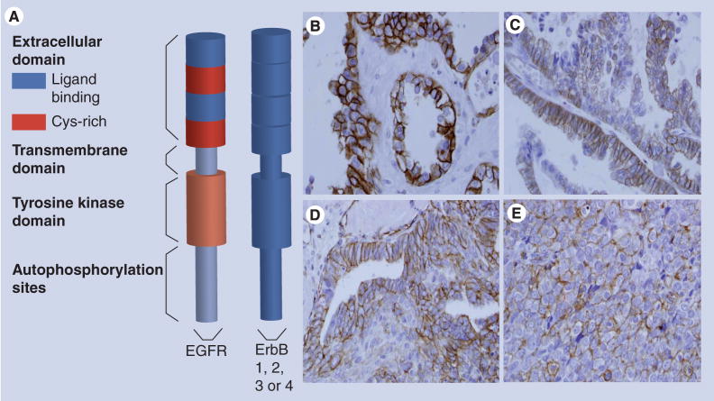
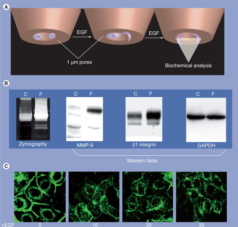
Ovarian carcinoma is most frequently detected when disease has already disseminated intra-abdominally, resulting in a 5-year survival rate of less than 20% owing to complications of metastasis. Peritoneal ascites is often present, establishing a unique microenvironmental niche comprised of tumor and inflammatory cells, along with a wide range of bioactive soluble factors, several of which stimulate the EGF-receptor (EGFR). Elevated EGFR is associated with less favorable disease outcome in ovarian cancer, related in part to EGFR activation of signaling cascades that lead to enhanced matrix metalloproteinase expression and/or function. The available data suggest that modulating the expression or activity of the EGFR and/or matrix metalloproteinases offers opportunity for targeted intervention in patients with metastatic disease.


Figures

Figure 1
Figure 1. Ovarian tumor microenvironment
(A) Malignant transformation and proliferation of OSE cells forms the primary ovarian tumor. (B) Ovarian cancer metastasis occurs by direct extension, via shedding of malignant cells into the peritoneal cavity. Exfoliated cells exist as MCAs or individual cells. Malignant ascites is common and is believed to promote dissemination of tumor cells intraperitoneally. (C) Intraperitoneal adhesion and localized invasion anchors secondary lesions in the peritoneum and omentum, followed by proliferation to establish disseminated intraperitoneal metastases. EMT: Epithelial–mesenchymal transition; MCA: Multicellular aggregates; OSE: Ovarian surface epithelium.
Figure 2
Figure 2. Activated EGF-receptor in ovarian tumors
(A) Model of the EGFR. The extracellular N-terminal domain contains two subdomains that directly interact with ligand and two cysteine-rich subdomains. There is a single transmembrane domain that links the extracellular domain to the intracellular tyrosine kinase domain and the C-terminal tail that contains the autophosphorylation sites. The EGFR dimerizes with other ErbB receptors. (B–E) Immunohistochemical staining for activated (phospho-)-EGFR in ovarian tumors. Immunohistochemical analysis was performed retrospectively as described in [41], using antibodies to phospho-EGFR (1:400, Zymed®, CA, USA) according to standard procedures. EGFR activation (phospho-EGFR) was evident in 35% of the specimens analyzed (n = 146) and MMP-9 expression was statistically positively correlated with EGFR activation [41]. (B) Ovarian clear cell carcinoma; (C) mucinous ovarian carcinoma; (D) endometrioid ovarian carcinoma; (E) serous ovarian carcinoma. EGFR: EGF-receptor.
Figure 3
Figure 3. MMP domain structure
(A) MT1-MMP (MMP-14) is a transmembrane protease comprised of a pro-peptide (processed intracellularly by furin in the secretory pathway; not shown) and a catalytic domain containing the Zn2+-binding consensus sequence HExxHxxGxxH. A flexible hinge region connects the catalytic domain to the hemopexin-like domain. The hemopexin-like domain may contain a dimerization interface and is important in substrate recognition. Following this region, a short stalk connects the transmembrane domain to the short cytoplasmic tail. In addition to degrading protein substrates such as interstitial (type I) collagen, MT1-MMP also catalyzes activation of pro-MMP-2. This reaction proceeds via an unusual mechanism requiring a trimeric complex among MT1-MMP, TIMP-2 and pro-MMP-2. Anchoring of pro-MMP-2 via trimeric complex formation enables cleavage of the pro-peptide domain of pro-MMP-2 by a second molecule of MT1-MMP, releasing soluble active MMP-2. (B) MMP-9 is initially secreted in zymogen form. Proteolytic processing of the pro-peptide region, catalyzed by numerous proteinases in the extracellular mileu, exposes the active Zn2+-binding catalytic domain. The active site of MMP-9 is also connected via a flexible hinge to a hemopexin-domain that imparts substrate specificity. MMP: Matrix metalloproteinase; MT1: Membrane type 1; TIMP-2: Tissue inhibitor of metalloproteinase-2.
Figure 4
Figure 4. EGF-receptor activation regulates matrix metalloproteinase localization
(A & B) Analysis of MMP-9 in filopodia of EGF-stimulated cells. OVCA429 cells (1 × 10⁶) were serum-starved overnight and plated onto six-well plates containing 1.0 μm pore size membrane inserts. Each bottom chamber contained 2.5 ml of serum-free medium containing EGF. Following a 12-h incubation (empirically determined based on the lack of visible nuclei on the filter bottom), lysates were individually collected from the top chamber (cellular fraction) or the bottom chamber (filopodial fraction). Lysates were pooled from replicate chambers, normalized for protein concentration, and an equal amount (40 μg) of filopodial or total cellular protein was evaluated by gelatin zymography or western blotting, as indicated. Note that MMP-9 protein and activity are enriched in the filopodial pool following EGF treatment. β1 integrin is also concentrated in filopodia, while GAPDH is evenly distributed. (C) Cells expressing GFP-tagged MT1-MMP were seeded onto 35 mm Becton glass bottom dishes and serum-starved overnight prior to treatment with EGF (25 nM). Images were acquired at the indicated time points using a Nikon TE2000 inverted microscope (Nikon Inc., NY, USA) with a 60× oil immersion objective with numerical aperture 1.40 and analyzed using Metamorph Software (Molecular Devices, PA, USA). Note that EGF induces rapid internalization of MT1-MMP. C: Cellular fraction; F: Filopodial fraction; GAPDH: Glyceraldehyde 3-phosphate dehydrogenase; GFP: Green fluorescent protein; MMP: Matrix metalloproteinase; MT1: Membrane type 1.

Similar articles

Cited by

References

Bibliography

    1. Shedden KA, Kshisager MP, Schwartz DR, et al. Histologic type, organ or origin and wnt pathway status: effect on gene expression in ovarian and uterine carcinomas. Clin Can Res. 2005;11:2123–2131. - PubMed
    1. Shih IM, Kurman RJ. Ovarian tumorigenesis: a proposed model based on morphological and molecular genetic analysis. Am J Path. 2004;164:1511–1518. - PMC - PubMed
    1. Schwartz DR, Kardia SLR, Shedden KA, et al. Gene expression in ovarian cancer reflects both morphology and biological behavior, distinguishing clear cell from other poor-prognosis ovarian carcinomas. Can Res. 2002;62:4722–4729. - PubMed
    1. Scully RE, Young RH, Clement PB. Atlas of Tumor Pathology (Third Series, Fascicle 23) Armed Forces Institute of Pathology; Washington, DC, USA: 1998. Tumors of the ovary, maldeveloped gonads, fallopian tube and broad ligament.
    1. Lafky JM, Wilken JA, Baron AT, Maihle NJ. Clinical implications of the ErbB/epidermal growth factor (EGF) receptor family and its ligands in ovarian cancer. Biochim Biophys Acta. 2008;1785(2):232–265. This article is an exceptionally thorough review of the literature on the expression of ErbB receptors and ligands and clinical correlates in ovarian cancer. - PubMed
Website
    1. National Cancer Institute. Surveillance Epidemiology and End Results (SEER) stat fact sheet . http://seer.cancer.gov/statfacts/html/ovary.html.

Publication types

MeSH terms