

Mitochondrial gene therapy augments mitochondrial physiology in a Parkinson's disease cell model
- PMID: 19374590
- PMCID: PMC2829286
- DOI: 10.1089/hum.2009.023
Mitochondrial gene therapy augments mitochondrial physiology in a Parkinson's disease cell model
Abstract
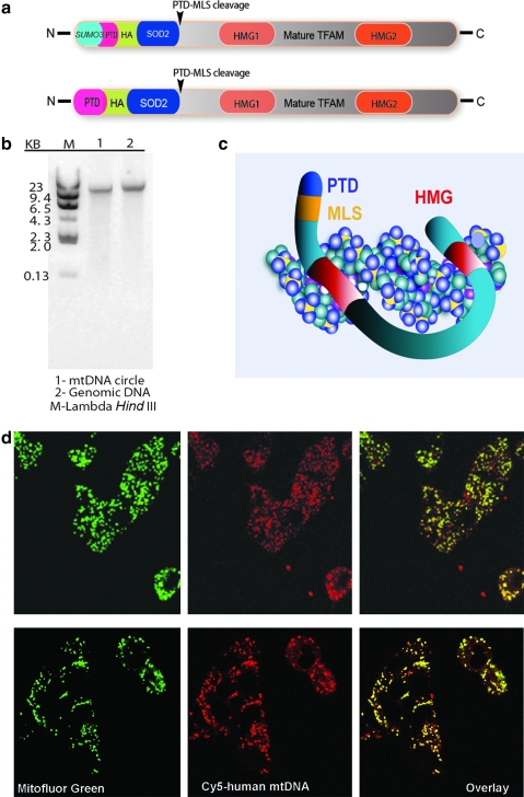
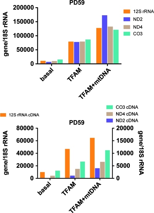
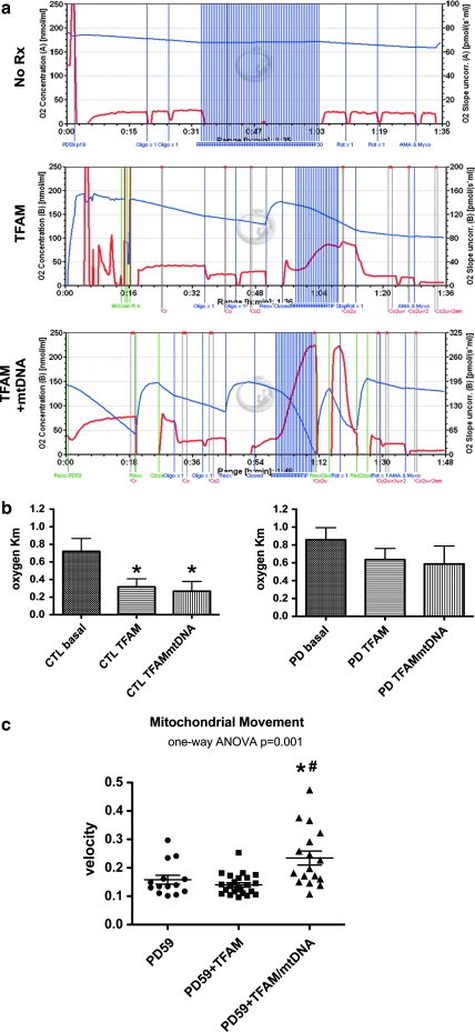
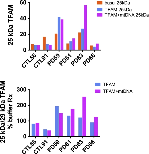
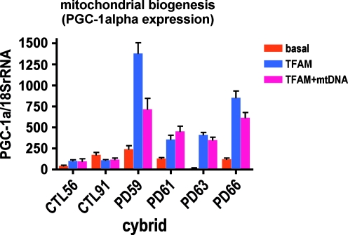
Neurodegeneration in Parkinson's disease (PD) affects mainly dopaminergic neurons in the substantia nigra, where age-related, increasing percentages of cells lose detectable respiratory activity associated with depletion of intact mitochondrial DNA (mtDNA). Replenishment of mtDNA might improve neuronal bioenergetic function and prevent further cell death. We developed a technology ("ProtoFection") that uses recombinant human mitochondrial transcription factor A (TFAM) engineered with an N-terminal protein transduction domain (PTD) followed by the SOD2 mitochondrial localization signal (MLS) to deliver mtDNA cargo to the mitochondria of living cells. MTD-TFAM (MTD = PTD + MLS = "mitochondrial transduction domain") binds mtDNA and rapidly transports it across plasma membranes to mitochondria. For therapeutic proof-of-principle we tested ProtoFection technology in Parkinson's disease cybrid cells, using mtDNA generated from commercially available human genomic DNA (gDNA; Roche). Nine to 11 weeks after single exposures to MTD-TFAM + mtDNA complex, PD cybrid cells with impaired respiration and reduced mtDNA genes increased their mtDNA gene copy numbers up to 24-fold, mtDNA-derived RNAs up to 35-fold, TFAM and ETC proteins, cell respiration, and mitochondrial movement velocities. Cybrid cells with no or minimal basal mitochondrial impairments showed reduced or no responses to treatment, suggesting the possibility of therapeutic selectivity. Exposure of PD but not control cybrid cells to MTD-TFAM protein alone or MTD-TFAM + mtDNA complex increased expression of PGC-1alpha, suggesting activation of mitochondrial biogenesis. ProtoFection technology for mitochondrial gene therapy holds promise for improving bioenergetic function in impaired PD neurons and needs additional development to define its pharmacodynamics and delineate its molecular mechanisms. It also is unclear whether single-donor gDNA for generating mtDNA would be a preferred therapeutic compared with the pooled gDNA used in this study.
Figures
Similar articles
-
Recombinant mitochondrial transcription factor A with N-terminal mitochondrial transduction domain increases respiration and mitochondrial gene expression.Mitochondrion. 2009 Jun;9(3):196-203. doi: 10.1016/j.mito.2009.01.012. Epub 2009 Feb 4. Mitochondrion. 2009. PMID: 19460293 Free PMC article.
-
Plausible Role of Mitochondrial DNA Copy Number in Neurodegeneration-a Need for Therapeutic Approach in Parkinson's Disease (PD).Mol Neurobiol. 2023 Dec;60(12):6992-7008. doi: 10.1007/s12035-023-03500-x. Epub 2023 Jul 31. Mol Neurobiol. 2023. PMID: 37523043 Review.
-
Cybrid models of Parkinson's disease show variable mitochondrial biogenesis and genotype-respiration relationships.Exp Neurol. 2009 Dec;220(2):374-82. doi: 10.1016/j.expneurol.2009.09.025. Epub 2009 Oct 6. Exp Neurol. 2009. PMID: 19815014 Free PMC article.
-
ERK-mediated phosphorylation of TFAM downregulates mitochondrial transcription: implications for Parkinson's disease.Mitochondrion. 2014 Jul;17:132-40. doi: 10.1016/j.mito.2014.04.008. Epub 2014 Apr 24. Mitochondrion. 2014. PMID: 24768991 Free PMC article.
-
Mitochondrial transcription factor A regulates mitochondrial transcription initiation, DNA packaging, and genome copy number.Biochim Biophys Acta. 2012 Sep-Oct;1819(9-10):921-9. doi: 10.1016/j.bbagrm.2012.03.002. Epub 2012 Mar 21. Biochim Biophys Acta. 2012. PMID: 22465614 Review.
Cited by
-
Comparison of three methods for mitochondria isolation from the human liver cell line (HepG2).Gastroenterol Hepatol Bed Bench. 2016 Spring;9(2):105-13. Gastroenterol Hepatol Bed Bench. 2016. PMID: 27099670 Free PMC article.
-
The Alzheimer's disease mitochondrial cascade hypothesis.J Alzheimers Dis. 2010;20 Suppl 2(Suppl 2):S265-79. doi: 10.3233/JAD-2010-100339. J Alzheimers Dis. 2010. PMID: 20442494 Free PMC article. Review.
-
LHON gene therapy vector prevents visual loss and optic neuropathy induced by G11778A mutant mitochondrial DNA: biodistribution and toxicology profile.Invest Ophthalmol Vis Sci. 2014 Oct 23;55(12):7739-53. doi: 10.1167/iovs.14-15388. Invest Ophthalmol Vis Sci. 2014. PMID: 25342621 Free PMC article.
-
Targeting Mitochondrial Impairment in Parkinson's Disease: Challenges and Opportunities.Front Cell Dev Biol. 2021 Jan 5;8:615461. doi: 10.3389/fcell.2020.615461. eCollection 2020. Front Cell Dev Biol. 2021. PMID: 33469539 Free PMC article. Review.
-
Aging-associated accumulation of mitochondrial DNA mutations in tumor origin.Life Med. 2022 Aug 17;1(2):149-167. doi: 10.1093/lifemedi/lnac014. eCollection 2022 Oct. Life Med. 2022. PMID: 39871923 Free PMC article. Review.
References
-
- Bannwarth S. Procaccio V. Paquis-Flucklinger V. Surveyor nuclease: A new strategy for a rapid identification of heteroplasmic mitochondrial DNA mutations in patients with respiratory chain defects. Hum. Mutat. 2005;25:575–582. - PubMed
-
- Bannwarth S. Procaccio V. Paquis-Flucklinger V. Rapid identification of unknown heteroplasmic mutations across the entire human mitochondrial genome with mismatch-specific Surveyor nuclease. Nat. Protoc. 2006;1:2037–2047. - PubMed
-
- Bender A. Krishnan K.J. Morris C.M. Taylor G.A. Reeve A.K. Perry R.H. Jaros E. Hersheson J.S. Betts J. Klopstock T. Taylor R.W. Turnbull D.M. High levels of mitochondrial DNA deletions in substantia nigra neurons in aging and Parkinson disease. Nat. Genet. 2006;38:515–517. - PubMed
-
- Bender A. Schwarzkopf R.M. McMillan A. Krishnan K.J. Rieder G. Neumann M. Elstner M. Turnbull D.M. Klopstock T. Dopaminergic midbrain neurons are the prime target for mitochondrial DNA deletions. J. Neurol. 2008;255:1231–1235. - PubMed
-
- Bogenhagen D.F. Rousseau D. Burke S. The layered structure of human mitochondrial DNA nucleoids. J. Biol. Chem. 2008;283:3665–3675. - PubMed
Publication types
MeSH terms
Substances
Grants and funding
LinkOut - more resources
Full Text Sources
Other Literature Sources
Medical
