. 2009 Apr 17:6:11.
doi: 10.1186/1743-8977-6-11.

Environmentally persistent free radicals amplify ultrafine particle mediated cellular oxidative stress and cytotoxicity

Affiliations

Environmentally persistent free radicals amplify ultrafine particle mediated cellular oxidative stress and cytotoxicity

Shrilatha Balakrishna et al. Part Fibre Toxicol. .

Abstract

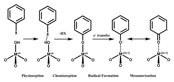
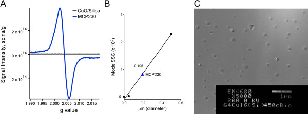
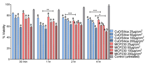
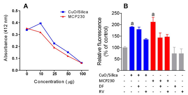
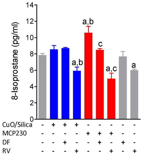
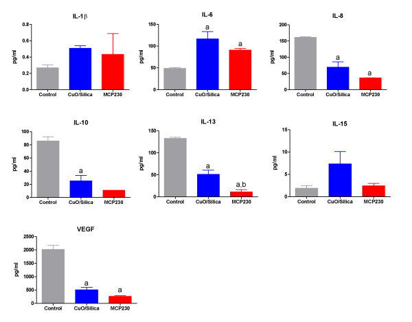
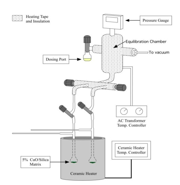
Background: Combustion generated particulate matter is deposited in the respiratory tract and pose a hazard to the lungs through their potential to cause oxidative stress and inflammation. We have previously shown that combustion of fuels and chlorinated hydrocarbons produce semiquinone-type radicals that are stabilized on particle surfaces (i.e. environmentally persistent free radicals; EPFRs). Because the composition and properties of actual combustion-generated particles are complex, heterogeneous in origin, and vary from day-to-day, we have chosen to use surrogate particle systems. In particular, we have chosen to use the radical of 2-monochlorophenol (MCP230) as the EPFR because we have previously shown that it forms a EPFR on Cu(II)O surfaces and catalyzes formation of PCDD/F. To understand the physicochemical properties responsible for the adverse pulmonary effects of combustion by-products, we have exposed human bronchial epithelial cells (BEAS-2B) to MCP230 or the CuO/silica substrate. Our general hypothesis was that the EPFR-containing particle would have greater toxicity than the substrate species.

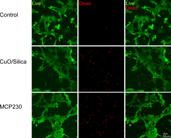
Results: Exposure of BEAS-2B cells to our combustion generated particle systems significantly increased reactive oxygen species (ROS) generation and decreased cellular antioxidants resulting in cell death. Resveratrol treatment reversed the decline in cellular glutathione (GSH), glutathione peroxidase (GPx), and superoxide dismutase (SOD) levels for both types of combustion-generated particle systems.

Conclusion: The enhanced cytotoxicity upon exposure to MCP230 correlated with its ability to generate more cellular oxidative stress and concurrently reduce the antioxidant defenses of the epithelial cells (i.e. reduced GSH, SOD activity, and GPx). The EPFRs in MCP230 also seem to be of greater biological concern due to their ability to induce lipid peroxidation. These results are consistent with the oxidizing nature of the CuO/silica ultrafine particles and the reducing nature and prolonged environmental and biological lifetimes of the EPFRs in MCP230.


Figures

Figure 1
Figure 1
Mechanism of formation of a phenoxyl-type PFR from a substituted aromatic on a metal oxide surface. PFR formation proceeds through a mechanism of: 1) physisorption, 2) chemisorption by elimination of HX, and electron transfer to form the surface-associated PFR and a reduced metal. The resulting radical may be primarily oxygen-centered or carbon-centered based on the properties of the PFR-metal complex.
Figure 2
Figure 2
Physical properties of ultrafine particles: (A) EPR spectra of CuO/Silica and CuO/Silica exposed to 2-monochlorophenol at 230°C (MCP230). (B) Data showing the calculated size of MCP230 by flow cytometry. (C) Transmission electron micrograph of 100–200 nm Cab-o-sil Silica particles, containing CuO nanoclusters in an isotonic saline solution containing 0.02% tween-80. The procedure followed in preparation of this particle suspension is exactly the same in all studies presented in this manuscript. This figure demonstrates that the particles exist as singlets without aggregation.
Figure 3
Figure 3
Cytotoxicity in BEAS-2B cells exposed to CuO/Silica or MCP230 ultrafine particles for 0.5, 1, 2 and 4 h. Results are expressed as mean ± SEM (n = 3). Significantly different from auntreated controls or bCuO/Silica at the same dose and exposure time (Two-way ANOVA; *P < 0.05, **P < 0.01, ***P < 0.001).
Figure 4
Figure 4
Cell-membrane integrity of BEAS-2B cells on exposure to CuO/Silica and MCP230 ultrafine particles. BEAS-2B cells were exposed for 4 h to 100 μg/cm2 ultrafine particles. After the exposure, cells were treated with calcein AM and ethidium homodimer-1. Calcein AM enters all cells and is enzymatically converted to calcein. Cells with an intact plasma membrane (viable cells) retain calcein in the cytoplasm and thus fluoresce green. Cells with a compromised plasma membrane (dead cells) take up ethidium homodimer-1, which fluoresces red when complexed with nucleic acid. Scale bar = 50 μm.
Figure 5
Figure 5
(A) DTT activity with CuO/Silica, and MCP230 ultrafine particles at various concentrations. (B) ROS generation in CuO/Silica or MCP230 ultrafine particles exposed BEAS-2B cells co-treated with 100 μM deferoxamine (DF) or 25 μM resveratrol (RV) for 4 h as measured by DCF assay. Results are expressed as mean ± SEM (n = 3). a significantly different from untreated controls.
Figure 6
Figure 6
Cellular antioxidant status of BEAS-2B cells exposed to CuO/Silica and MCP230 ultrafine particles. Cells were co-treated with 100 μM deferoxamine (DF) or 25 μM resveratrol (RV) ± CuO/or MCP230 ultrafine particles for 4 h. (A) Depletion of intracellular glutathione (GSH) (B) Cytosolic superoxide dismutase (SOD) activity (C) Alterations in the glutathione peroxidase (GPx) enzyme activity. Results are expressed as mean ± SEM (n = 3). Significantly different from auntreated controls; bCuO/Silica; cMCP230.
Figure 7
Figure 7
Levels of by 8-isoprostane in BEAS -2B cell supernatant. BEAS-2B cells were co-treated with 100 μM deferoxamine or 25 μM resveratrol and CuO/Silica or MCP230 ultrafine particles for 4 h in a 6-well plate with 1 ml culture media. Results are expressed as mean ± SEM. Significantly different from auntreated controls; bCuO/Silica; cMCP230.
Figure 8
Figure 8
Effect of CuO/Silica and MCP ultrafine particles on cytokine release from BEAS-2B at 4 h post-exposure. Since IL-2, IL-12p40, IL12p70, IL-18, IFNγ, TNFα, IFNα2 were not detected at 4 h after ultrafine particle exposure, they were excluded from the figure. Results are expressed as mean ± SEM (n = 3). Significantly different from auntreated controls, bbetween CuO/Silica and MCP230 groups.
Figure 9
Figure 9
Pictorial diagram of a two-port, particle dosing apparatus.

References

    1. Van Eeden SF, Yeung A, Quinlam K, Hogg JC. Systemic response to ambient particulate matter: relevance to chronic obstructive pulmonary disease. Proc Am Thorac Soc. 2005;2:61–67. doi: 10.1513/pats.200406-035MS. - DOI - PubMed
    1. Hamade AK, Rabold R, Tankersley CG. Adverse cardiovascular effects with acute particulate matter and ozone exposures: interstrain variation in mice. Environ Health Perspect. 2008;116:1033–1039. - PMC - PubMed
    1. Arden Pope C, Burnett RT, Thun MJ, Calle EE, Krewski D, Ito K, Thurston GD. Lung Cancer, Cardiopulmonary Mortality, and Long-term Exposure to Fine Particulate Air Pollution. JAMA. 2002;287:1132–1141. doi: 10.1001/jama.287.9.1132. - DOI - PMC - PubMed
    1. Avakian MD, Dellinger B, Fiedler H, Gullet B, Koshland C, Marklund S, Oberdörster G, Safe S, Sarofim A, Smith KR, Schwartz D, Suk WA. The origin, fate, and health effects of combustion by-products: a research framework. Environ Health Perspect. 2002;110:1155–1162. - PMC - PubMed
    1. Cormier SA, Lomnicki S, Backes W, Dellinger B. Origin and health impacts of emissions of toxic by-products and fine particles from combustion and thermal treatment of hazardous wastes and materials. Environ Health Perspect. 2006;114:810–817. - PMC - PubMed

LinkOut - more resources