

Diet-induced obesity causes cerebral vessel remodeling and increases the damage caused by ischemic stroke
- PMID: 19374911
- PMCID: PMC3959658
- DOI: 10.1016/j.mvr.2009.04.004
Diet-induced obesity causes cerebral vessel remodeling and increases the damage caused by ischemic stroke
Abstract
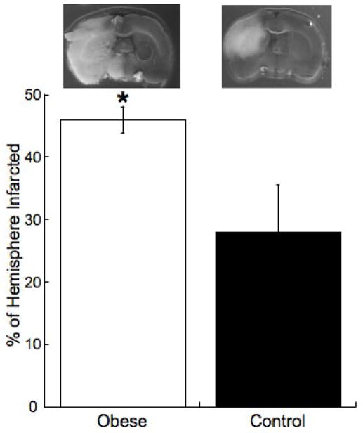
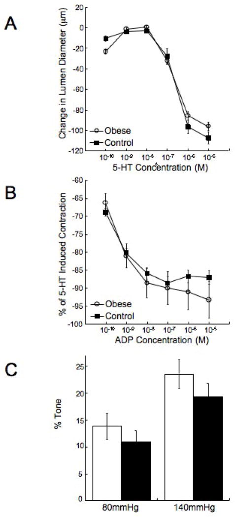
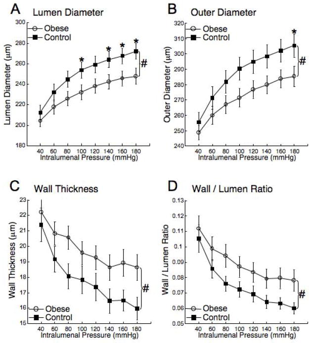
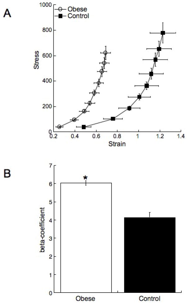
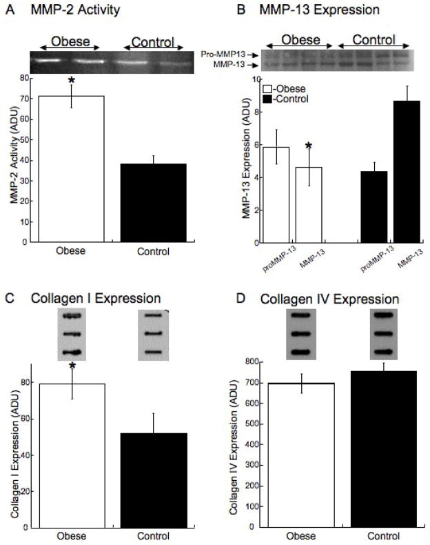
Hypertension, elevated fasting blood glucose and plasma insulin develop in rats fed a high fat (HF) diet. Our goal was to assess the effects of obesity, beginning in childhood, on the adult cardiovascular system. We hypothesized that rats fed a HF diet would have larger ischemic cerebral infarcts and middle cerebral artery (MCA) remodeling. Three-week-old male Sprague Dawley rats were fed a HF (obese) or control diet for 10 weeks. Cerebral ischemia was induced by MCA occlusion (MCAO). MCA structure was assessed by pressure myography and cerebral vessel matrix metalloproteinase (MMP) activity and expression and collagen levels were measured in vessels from rats that did not undergo MCAO. The cerebral infarct was greater in the obese rats than the control (46.0+/-2.1 vs 28.0+/-7.5% of the hemisphere infarcted, obese vs control p<0.05). The MCAs from obese rats had smaller lumens (232+/-7.2 vs 254+/-7.8 microm obese vs control p<0.05) and thicker walls (19.6+/-0.8 vs 17.8+/-0.9 microm obese vs control p<0.05) and were less compliant than MCAs from control rats. MMP-2 activity and collagen I expression were increased in vessels from obese rats and MMP-13 expression was reduced. These results suggest that obesity, beginning in childhood, causes inward vessel remodeling with a concomitant increase in vessel stiffness due to increased collagen deposition. These changes in MCA structure may be responsible for the increase in the ischemic damage after MCAO.
Figures
Similar articles
-
Mineralocorticoid receptor antagonism prevents obesity-induced cerebral artery remodeling and reduces white matter injury in rats.Microcirculation. 2018 Jul;25(5):e12460. doi: 10.1111/micc.12460. Epub 2018 Jun 7. Microcirculation. 2018. PMID: 29758591 Free PMC article.
-
Obesity increases blood pressure, cerebral vascular remodeling, and severity of stroke in the Zucker rat.Hypertension. 2009 Feb;53(2):381-6. doi: 10.1161/HYPERTENSIONAHA.108.124149. Epub 2008 Dec 22. Hypertension. 2009. PMID: 19104000 Free PMC article.
-
11beta-hydroxysteroid dehydrogenase type II inhibition causes cerebrovascular remodeling and increases infarct size after cerebral ischemia.Endocrinology. 2009 Feb;150(2):713-9. doi: 10.1210/en.2008-0808. Epub 2008 Oct 9. Endocrinology. 2009. PMID: 18845645 Free PMC article.
-
Regional cerebral blood flow after occlusion of the middle cerebral artery.Acta Neurol Scand. 1986 Apr;73(4):321-37. doi: 10.1111/j.1600-0404.1986.tb03286.x. Acta Neurol Scand. 1986. PMID: 3524091 Review.
-
The effects of obesity on the cerebral vasculature.Curr Vasc Pharmacol. 2014 May;12(3):462-72. doi: 10.2174/1570161112666140423222411. Curr Vasc Pharmacol. 2014. PMID: 24846235 Free PMC article. Review.
Cited by
-
A 2-Min Transient Ischemia Confers Cerebral Ischemic Tolerance in Non-Obese Gerbils, but Results in Neuronal Death in Obese Gerbils by Increasing Abnormal mTOR Activation-Mediated Oxidative Stress and Neuroinflammation.Cells. 2019 Sep 22;8(10):1126. doi: 10.3390/cells8101126. Cells. 2019. PMID: 31546722 Free PMC article.
-
How Brain Infarction Links With the Microbiota-Gut-Brain Axis: Hints From Studies Focusing on the Risk Factors for Ischemic Stroke.Front Neurosci. 2022 May 24;16:877937. doi: 10.3389/fnins.2022.877937. eCollection 2022. Front Neurosci. 2022. PMID: 35685776 Free PMC article. Review.
-
Mineralocorticoid receptor antagonism prevents obesity-induced cerebral artery remodeling and reduces white matter injury in rats.Microcirculation. 2018 Jul;25(5):e12460. doi: 10.1111/micc.12460. Epub 2018 Jun 7. Microcirculation. 2018. PMID: 29758591 Free PMC article.
-
Obesity and stroke: Can we translate from rodents to patients?J Cereb Blood Flow Metab. 2016 Dec;36(12):2007-2021. doi: 10.1177/0271678X16670411. Epub 2016 Sep 21. J Cereb Blood Flow Metab. 2016. PMID: 27655337 Free PMC article. Review.
-
High-fat diet increases O-GlcNAc levels in cerebral arteries: a link to vascular dysfunction associated with hyperlipidaemia/obesity?Clin Sci (Lond). 2016 Jun 1;130(11):871-80. doi: 10.1042/CS20150777. Epub 2016 Feb 29. Clin Sci (Lond). 2016. PMID: 26929437 Free PMC article.
References
-
- Bagust A, et al. The projected health care burden of Type 2 diabetes in the UK from 2000 to 2060. Diabet Med. 2002;19(Suppl 4):1–5. - PubMed
-
- Baumbach GL, Heistad DD. Remodeling of cerebral arterioles in chronic hypertension. Hypertension. 1989;13:968–72. - PubMed
-
- Bouvet C, et al. Different involvement of extracellular matrix components in small and large arteries during chronic NO synthase inhibition. Hypertension. 2005;45:432–7. - PubMed
-
- Carroll JF, et al. Hypertension, cardiac hypertrophy, and neurohumoral activity in a new animal model of obesity. Am J Physiol. 1996;271:H373–8. - PubMed
-
- Cipolla MJ, et al. Postischemic attenuation of cerebral artery reactivity is increased in the presence of tissue plasminogen activator. Stroke. 2000;31:940–5. - PubMed
Publication types
MeSH terms
Substances
Grants and funding
LinkOut - more resources
Full Text Sources
Medical
Research Materials
Miscellaneous
