

Striatal dopamine and glutamate receptors modulate methamphetamine-induced cortical Fos expression
- PMID: 19374938
- PMCID: PMC2716135
- DOI: 10.1016/j.neuroscience.2009.04.023
Striatal dopamine and glutamate receptors modulate methamphetamine-induced cortical Fos expression
Abstract
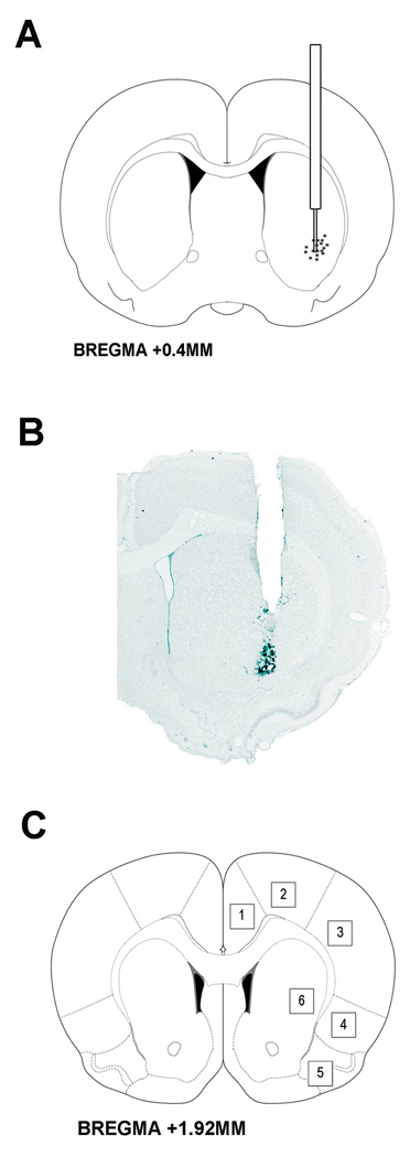
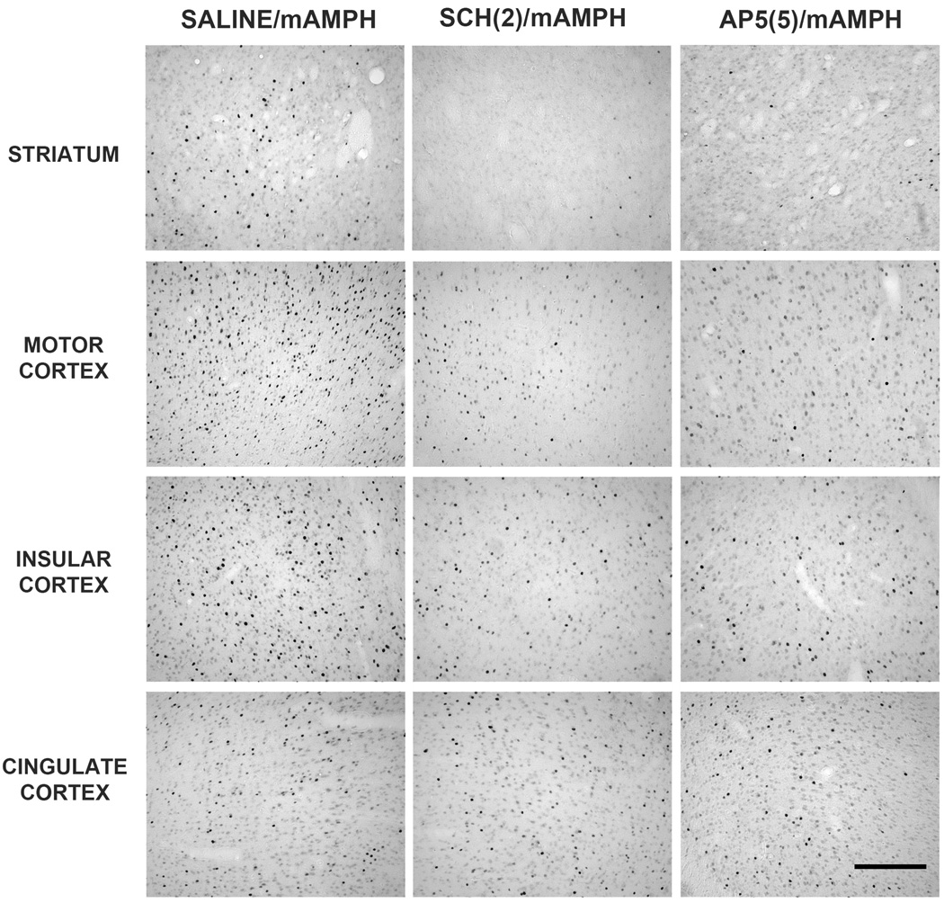
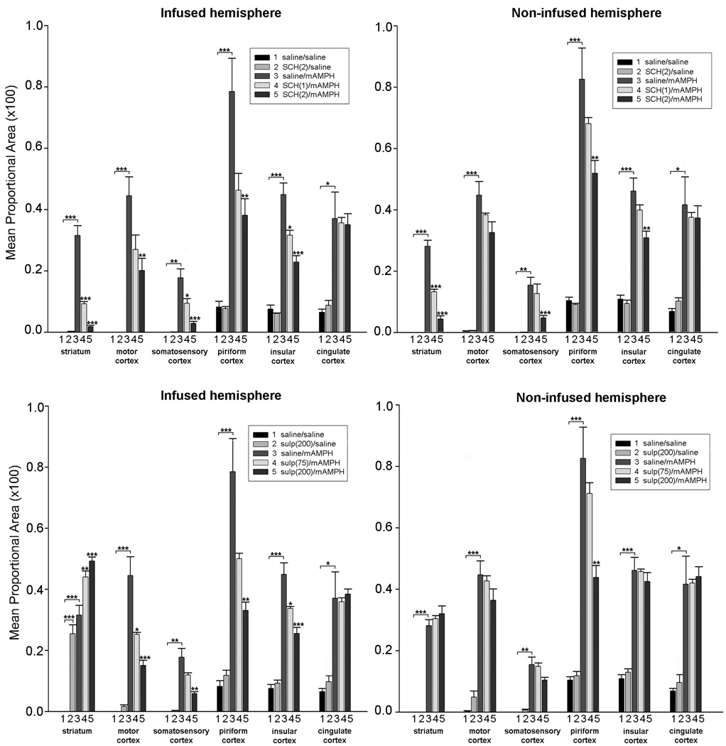
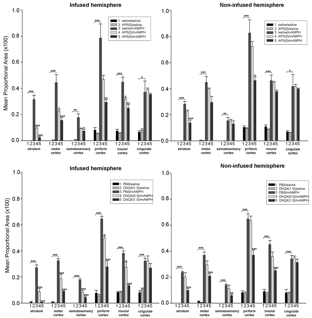
Methamphetamine (mAMPH) is a psychostimulant drug that increases extracellular levels of monoamines throughout the brain. It has previously been observed that a single injection of mAMPH increases immediate early gene (IEG) expression in both the striatum and cerebral cortex. Moreover, this effect is modulated by dopamine and glutamate receptors since systemic administration of dopamine or glutamate antagonists has been found to alter mAMPH-induced striatal and cortical IEG expression. However, because dopamine and glutamate receptors are found in extra-striatal as well as striatal brain regions, studies employing systemic injection of dopamine or glutamate antagonists fail to localize the effects of mAMPH-induced activation. In the present experiments, the roles of striatal dopamine and glutamate receptors in mAMPH-induced gene expression in the striatum and cerebral cortex were examined. The nuclear expression of Fos, the protein product of the IEG c-fos, was quantified in both the striatum and the cortex of animals receiving intrastriatal dopamine or glutamate antagonist administration. Intrastriatal infusion of dopamine (D1 or D2) or glutamate [N-methyl-D-aspartic acid (NMDA) or alpha-amino-3-hydroxy-5-methyl-4-isoxazolepropionic acid (AMPA)] antagonists affected not only mAMPH-induced striatal, but also cortical, Fos expression. Overall, the effects of the antagonists occurred dose-dependently, in both the infused and non-infused hemispheres, with greater influences occurring in the infused hemisphere. Finally, unilateral intrastriatal infusion of dopamine or glutamate antagonists changed the behavior of the rats from characteristic mAMPH-induced stereotypy to rotation ipsilateral to the infusion. These results demonstrate that mAMPH's actions on striatal dopamine and glutamate receptors modulate the widespread cortical activation induced by mAMPH. It is hypothesized that dopamine release from nigrostriatal terminals modulates activity within striatal efferent pathways, thereby disinhibiting thalamo-cortical circuits. By extension, these results suggest processes through which repeated exposure to mAMPH might influence cortical function in mAMPH abusers.
Figures
Similar articles
-
Cortical ionotropic glutamate receptor antagonism protects against methamphetamine-induced striatal neurotoxicity.Neuroscience. 2011 Dec 29;199:272-83. doi: 10.1016/j.neuroscience.2011.09.014. Epub 2011 Sep 16. Neuroscience. 2011. PMID: 21946008 Free PMC article.
-
Striatal dopamine D1 and D2 receptors: widespread influences on methamphetamine-induced dopamine and serotonin neurotoxicity.Synapse. 2011 Nov;65(11):1144-55. doi: 10.1002/syn.20952. Epub 2011 Aug 31. Synapse. 2011. PMID: 21584865
-
Cortical stimulation induces Fos expression in striatal neurons via NMDA glutamate and dopamine receptors.Brain Res. 1995 Nov 27;700(1-2):1-12. doi: 10.1016/0006-8993(95)00958-s. Brain Res. 1995. PMID: 8624698
-
NMDA and D1 receptors mediate induction of c-fos and junB genes in striatum following morphine administration: implications for studies of memory.Behav Brain Res. 1995 Jan 23;66(1-2):225-30. doi: 10.1016/0166-4328(94)00146-7. Behav Brain Res. 1995. PMID: 7755894 Review.
-
The effect of striatal dopamine depletion on striatal and cortical glutamate: A mini-review.Prog Neuropsychopharmacol Biol Psychiatry. 2016 Feb 4;65:49-53. doi: 10.1016/j.pnpbp.2015.08.013. Epub 2015 Sep 1. Prog Neuropsychopharmacol Biol Psychiatry. 2016. PMID: 26334687 Free PMC article. Review.
Cited by
-
Addiction-related gene regulation: risks of exposure to cognitive enhancers vs. other psychostimulants.Prog Neurobiol. 2013 Jan;100:60-80. doi: 10.1016/j.pneurobio.2012.10.001. Epub 2012 Oct 17. Prog Neurobiol. 2013. PMID: 23085425 Free PMC article. Review.
-
Striatal patch compartment lesions alter methamphetamine-induced behavior and immediate early gene expression in the striatum, substantia nigra and frontal cortex.Brain Struct Funct. 2014 Jul;219(4):1213-29. doi: 10.1007/s00429-013-0559-x. Epub 2013 Apr 27. Brain Struct Funct. 2014. PMID: 23625147 Free PMC article.
-
Effects of low-doses of methamphetamine on d-fenfluramine-induced head-twitch response (HTR) in mice during ageing and c-fos expression in the prefrontal cortex.BMC Neurosci. 2023 Jan 11;24(1):2. doi: 10.1186/s12868-022-00766-0. BMC Neurosci. 2023. PMID: 36631757 Free PMC article.
-
Effects of intrastriatal dopamine D1 or D2 antagonists on methamphetamine-induced egocentric and allocentric learning and memory deficits in Sprague-Dawley rats.Psychopharmacology (Berl). 2019 Jul;236(7):2243-2258. doi: 10.1007/s00213-019-05221-3. Epub 2019 Mar 27. Psychopharmacology (Berl). 2019. PMID: 30919007 Free PMC article.
-
Modulation of dlPFC function and decision-making capacity by repetitive transcranial magnetic stimulation in methamphetamine use disorder.Transl Psychiatry. 2024 Jul 8;14(1):280. doi: 10.1038/s41398-024-03000-z. Transl Psychiatry. 2024. PMID: 38977700 Free PMC article.
References
-
- Albin RL, Makowiec RL, Hollingsworth ZR, Dure LSt, Penney JB, Young AB. Excitatory amino acid binding sites in the basal ganglia of the rat: a quantitative autoradiographic study. Neuroscience. 1992;46:35–48. - PubMed
-
- Alburges ME, Hunt ME, McQuade RD, Wamsley JK. D1-receptor antagonists: comparison of [3H]SCH39166 to [3H]SCH23390. J Chem Neuroanat. 1992;5:357–366. - PubMed
-
- Alexander GE, Crutcher MD. Functional architecture of basal ganglia circuits: neural substrates of parallel processing. Trends Neurosci. 1990;13:266–271. - PubMed
-
- Altar CA, O'Neil S, Walter RJ, Jr, Marshall JF. Brain dopamine and serotonin receptor sites revealed by digital subtraction autoradiography. Science. 1985;228:597–600. - PubMed
-
- Badiani A, Oates MM, Day HE, Watson SJ, Akil H, Robinson TE. Environmental modulation of amphetamine-induced c-fos expression in D1 versus D2 striatal neurons. Behav Brain Res. 1999;103:203–209. - PubMed
Publication types
MeSH terms
Substances
Grants and funding
LinkOut - more resources
Full Text Sources
Medical
