

Spatial and intracellular relationships between the alpha7 nicotinic acetylcholine receptor and the vesicular acetylcholine transporter in the prefrontal cortex of rat and mouse
- PMID: 19374941
- PMCID: PMC2720620
- DOI: 10.1016/j.neuroscience.2009.04.024
Spatial and intracellular relationships between the alpha7 nicotinic acetylcholine receptor and the vesicular acetylcholine transporter in the prefrontal cortex of rat and mouse
Abstract
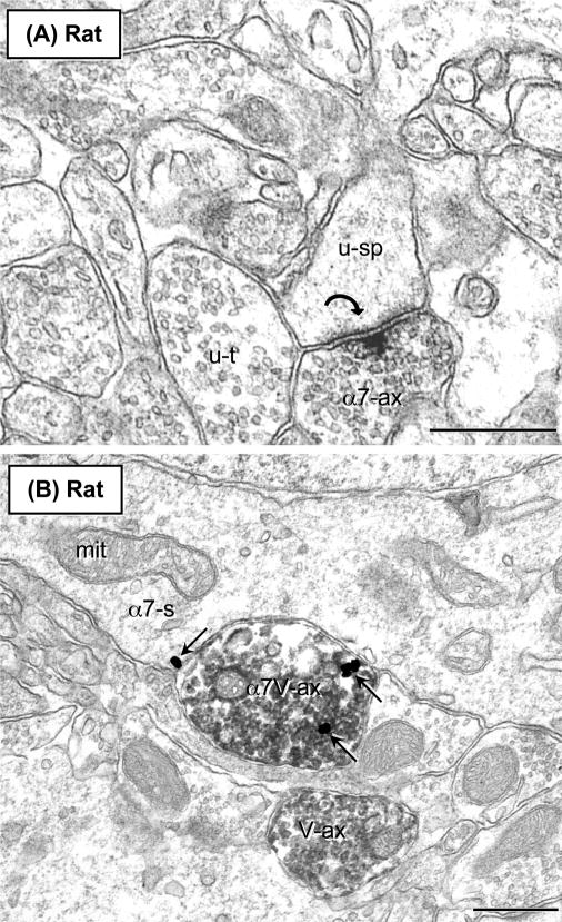
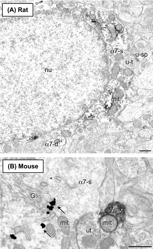
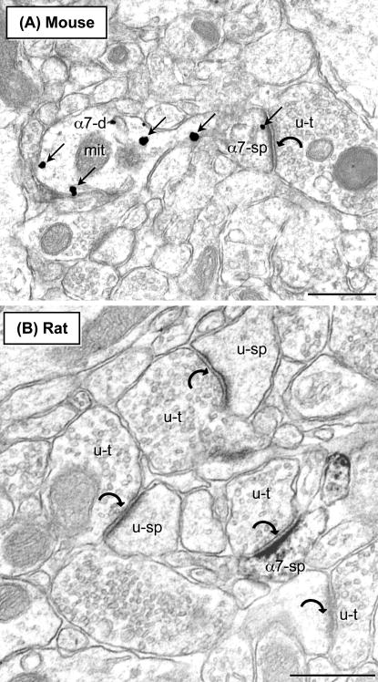
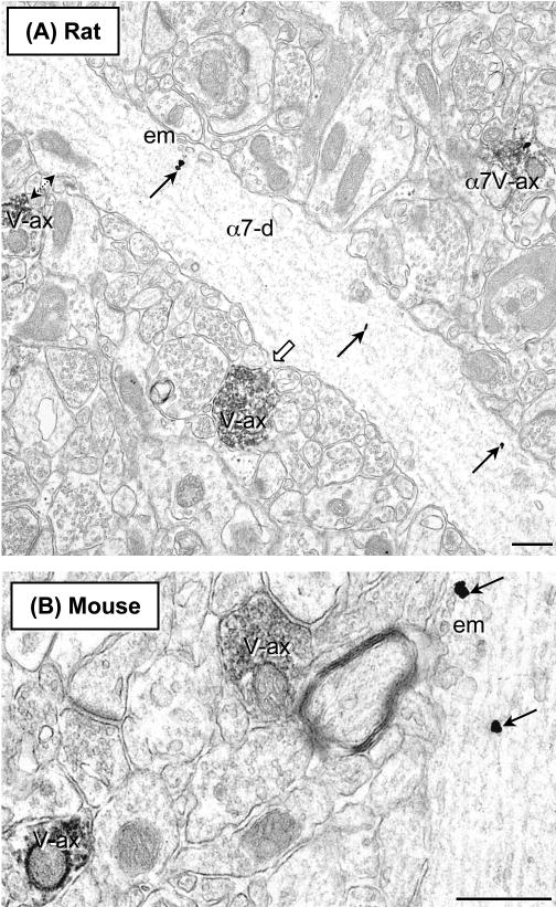
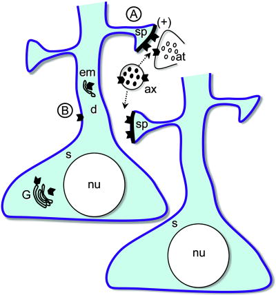
The alpha 7 subunit of the nicotinic acetylcholine receptor (alpha7nAChR) is expressed in the prefrontal cortex (PFC), a brain region where these receptors are implicated in cognitive function and in the pathophysiology of schizophrenia. Activation of this receptor is dependent on release of acetylcholine (ACh) from axon terminals that contain the vesicular acetylcholine transporter (VAChT). Since rat and mouse models are widely used for studies of specific abnormalities in schizophrenia, we sought to determine the subcellular location of the alpha7nAChR with respect to VAChT storage vesicles in axon terminals in the PFC in both species. For this, we used dual electron microscopic immunogold and immunoperoxidase labeling of antisera raised against the alpha7nAChR and VAChT. In both species, the alpha7nAChR-immunoreactivity ((-)ir) was principally identified within dendrites and dendritic spines, receptive to axon terminals forming asymmetric excitatory-type synapses, but lacking detectable alpha7nAChR or VAChT-ir. Quantitative analysis of the rat PFC revealed that of alpha7nAChR-labeled neuronal profiles, 65% (299/463) were postsynaptic structures (dendrites and dendritic spine) and only 22% (104/463) were axon terminals or small unmyelinated axons. In contrast, VAChT was principally localized to varicose vesicle-filled axonal profiles, without recognized synaptic specializations (n=240). Of the alpha7nAChR-labeled axons, 47% (37/79) also contained VAChT, suggesting that ACh release is autoregulated through the presynaptic alpha7nAChR. The VAChT-labeled terminals rarely formed synapses, but frequently apposed alpha7nAChR-containing neuronal profiles. These results suggest that in rodent PFC, the alpha7nAChR plays a major role in modulation of the postsynaptic excitation in spiny dendrites in contact with VAChT containing axons.
Figures
Similar articles
-
Acetylcholine α7 nicotinic and dopamine D2 receptors are targeted to many of the same postsynaptic dendrites and astrocytes in the rodent prefrontal cortex.Synapse. 2011 Dec;65(12):1350-67. doi: 10.1002/syn.20977. Synapse. 2011. PMID: 21858872 Free PMC article.
-
Dendritic and axonal targeting of the vesicular acetylcholine transporter to membranous cytoplasmic organelles in laterodorsal and pedunculopontine tegmental nuclei.J Comp Neurol. 2000 Mar 27;419(1):32-48. doi: 10.1002/(sici)1096-9861(20000327)419:1<32::aid-cne2>3.0.co;2-o. J Comp Neurol. 2000. PMID: 10717638
-
Muscarinic cholinergic receptor M1 in the rat basolateral amygdala: ultrastructural localization and synaptic relationships to cholinergic axons.J Comp Neurol. 2013 Jun 1;521(8):1743-59. doi: 10.1002/cne.23254. J Comp Neurol. 2013. PMID: 23559406 Free PMC article.
-
Cholinergic axon terminals in the ventral tegmental area target a subpopulation of neurons expressing low levels of the dopamine transporter.J Comp Neurol. 1999 Jul 26;410(2):197-210. doi: 10.1002/(sici)1096-9861(19990726)410:2<197::aid-cne3>3.0.co;2-d. J Comp Neurol. 1999. PMID: 10414527 Review.
-
Electron microscopic immunolabeling of transporters and receptors identifies transmitter-specific functional sites envisioned in Cajal's neuron.Prog Brain Res. 2002;136:145-55. doi: 10.1016/s0079-6123(02)36014-x. Prog Brain Res. 2002. PMID: 12143378 Review.
Cited by
-
α7 nicotinic ACh receptor-deficient mice exhibit sustained attention impairments that are reversed by β2 nicotinic ACh receptor activation.Br J Pharmacol. 2015 Oct;172(20):4919-31. doi: 10.1111/bph.13260. Epub 2015 Sep 22. Br J Pharmacol. 2015. PMID: 26222090 Free PMC article.
-
Dopamine receptor signaling in the medial orbital frontal cortex and the acquisition and expression of fructose-conditioned flavor preferences in rats.Brain Res. 2015 Jan 30;1596:116-25. doi: 10.1016/j.brainres.2014.11.028. Epub 2014 Nov 20. Brain Res. 2015. PMID: 25446441 Free PMC article.
-
Cancer 'survivor-care': I. the α7 nAChR as potential target for chemotherapy-related cognitive impairment.J Clin Pharm Ther. 2011 Aug;36(4):437-45. doi: 10.1111/j.1365-2710.2010.01208.x. Epub 2010 Aug 24. J Clin Pharm Ther. 2011. PMID: 21729110 Free PMC article. Review.
-
Basal Forebrain Cholinergic Circuits and Signaling in Cognition and Cognitive Decline.Neuron. 2016 Sep 21;91(6):1199-1218. doi: 10.1016/j.neuron.2016.09.006. Neuron. 2016. PMID: 27657448 Free PMC article. Review.
-
Neuromodulation of thought: flexibilities and vulnerabilities in prefrontal cortical network synapses.Neuron. 2012 Oct 4;76(1):223-39. doi: 10.1016/j.neuron.2012.08.038. Neuron. 2012. PMID: 23040817 Free PMC article. Review.
References
-
- Alkondon M, Rocha E, Maelicke A, Albuquerque E. Diversity of nicotinic acetylcholine receptors in rat brain. V. alpha-bungarotoxin-sensitive nicotinic receptors in olfactory bulb neurons and presynaptic modulation of glutamate release. J Pharmacol Exp Ther. 1996;278:1460–1471. - PubMed
-
- Araujo DM, Lapchak PA, Collier B, Quirion R. Characterization of N-[3H]methylcarbamylcholine binding sites and effect of N-methylcarbamylcholine on acetylcholine release in rat brain. J Neurochem. 1988;51:292–299. - PubMed
-
- Arnaiz-Cot JJ, Gonzalez JC, Sobrado M, Baldelli P, Carbone E, Gandia L, Garcia AG, Hernandez-Guijo JM. Allosteric modulation of α7 nicotinic receptors selectively depolarizes hippocampal interneurons, enhancing spontaneous GABAergic transmission. Eur J Neurosci. 2008;27:1097–1110. - PubMed
-
- Arvidsson U, Ried M, Elde R, Meister B. Vesicular acetylcholine transporter (VAChT) protein: A novel and unique marker for cholinergic neurons in the central and peripheral nervous systems. J Comp Neurol. 1997;378:454–467. - PubMed
-
- Barik J, Wonnacott S. Indirect modulation by α7 nicotinic acetylcholine receptors of noradrenaline release in rat hippocampal slices: Interaction with glutamate and GABA systems and effect of nicotine withdrawal. Mol Pharmacol. 2006;69:618–628. - PubMed
Publication types
MeSH terms
Substances
Grants and funding
LinkOut - more resources
Full Text Sources
Molecular Biology Databases
Miscellaneous
