

Impact of atypical retardation patterns on detection of glaucoma progression using the GDx with variable corneal compensation
- PMID: 19375062
- PMCID: PMC2848161
- DOI: 10.1016/j.ajo.2009.01.021
Impact of atypical retardation patterns on detection of glaucoma progression using the GDx with variable corneal compensation
Abstract
Purpose: To evaluate the impact of atypical retardation patterns (ARP) on detection of progressive retinal nerve fiber layer (RNFL) loss using scanning laser polarimetry with variable corneal compensation (VCC).
Design: Observational cohort study.
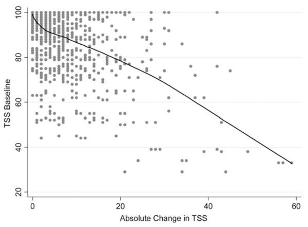
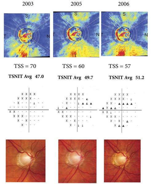
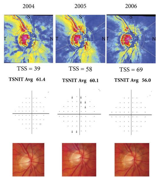
Methods: The study included 377 eyes of 221 patients with a median follow-up of 4.0 years. Images were obtained annually with the GDx VCC (Carl Zeiss Meditec Inc, Dublin, California, USA), along with optic disc stereophotographs and standard automated perimetry (SAP) visual fields. Progression was determined by the Guided Progression Analysis software for SAP and by masked assessment of stereophotographs by expert graders. The typical scan score (TSS) was used to quantify the presence of ARPs on GDx VCC images. Random coefficients models were used to evaluate the relationship between ARP and RNFL thickness measurements over time.
Results: Thirty-eight eyes (10%) showed progression over time on visual fields, stereophotographs, or both. Changes in TSS scores from baseline were significantly associated with changes in RNFL thickness measurements in both progressing and nonprogressing eyes. Each 1-unit increase in TSS score was associated with a 0.19-microm decrease in RNFL thickness measurement (P < .001) over time.
Conclusions: ARPs had a significant effect on detection of progressive RNFL loss with the GDx VCC. Eyes with large amounts of atypical patterns, great fluctuations on these patterns over time, or both may show changes in measurements that can appear falsely as glaucomatous progression or can mask true changes in the RNFL.
Figures

References
-
- Weinreb RN, Dreher AW, Coleman A, et al. Histopathologic validation of Fourier-ellipsometry measurements of retinal nerve fiber layer thickness. Arch Ophthalmol. 1990;108:557–560. - PubMed
-
- Zhou Q, Weinreb RN. Individualized compensation of anterior segment birefringence during scanning laser polarimetry. Invest Ophthalmol Vis Sci. 2002;43:2221–2228. - PubMed
-
- Weinreb RN, Bowd C, Zangwill LM. Glaucoma detection using scanning laser polarimetry with variable corneal polarization compensation. Arch Ophthalmol. 2003;121:218–224. - PubMed
-
- Greenfield DS, Knighton RW, Feuer WJ, Schiffman JC. Normative retardation data corrected for the corneal polarization axis with scanning laser polarimetry. Ophthalmic Surg Lasers Imaging. 2003;34:165–171. - PubMed
-
- Medeiros FA, Zangwill LM, Bowd C, et al. Fourier analysis of scanning laser polarimetry measurements with variable corneal compensation in glaucoma. Invest Ophthalmol Vis Sci. 2003;44:2606–2612. - PubMed
Publication types
MeSH terms
Grants and funding
LinkOut - more resources
Full Text Sources
Medical
Miscellaneous
