. 2009 May 12;19(9):723-9.
doi: 10.1016/j.cub.2009.03.056. Epub 2009 Apr 16.

aPKC phosphorylates Miranda to polarize fate determinants during neuroblast asymmetric cell division

Affiliations

aPKC phosphorylates Miranda to polarize fate determinants during neuroblast asymmetric cell division

Scott X Atwood et al. Curr Biol. .

Abstract

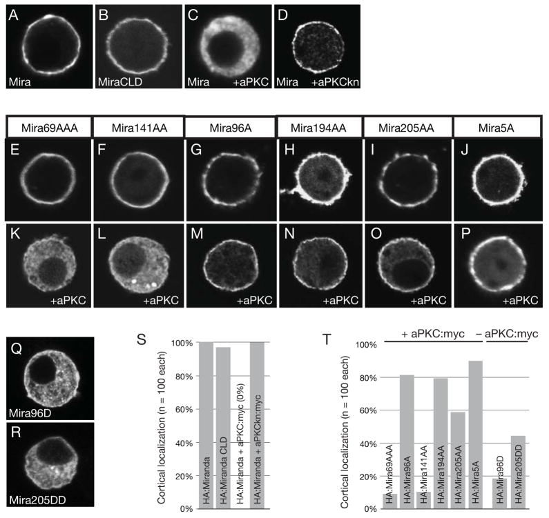
Background: Asymmetric cell divisions generate daughter cells with distinct fates by polarizing fate determinants into separate cortical domains. Atypical protein kinase C (aPKC) is an evolutionarily conserved regulator of cell polarity. In Drosophila neuroblasts, apically restricted aPKC is required for segregation of neuronal differentiation factors such as Numb and Miranda to the basal cortical domain. Whereas Numb is polarized by direct aPKC phosphorylation, Miranda asymmetry is thought to occur via a complicated cascade of repressive interactions (aPKC -| Lgl -| myosin II -| Miranda).

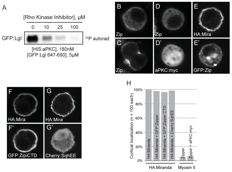
Results: Here we provide biochemical, cellular, and genetic data showing that aPKC directly phosphorylates Miranda to exclude it from the cortex and that Lgl antagonizes this activity. Miranda is phosphorylated by aPKC at several sites in its cortical localization domain and phosphorylation is necessary and sufficient for cortical displacement, suggesting that the repressive-cascade model is incorrect. In investigating key results that led to this model, we found that Y-27632, a Rho kinase inhibitor used to implicate myosin II, efficiently inhibits aPKC. Lgl3A, a nonphosphorylatable Lgl variant used to implicate Lgl in this process, inhibits the formation of apical aPKC crescents in neuroblasts. Furthermore, Lgl directly inhibits aPKC kinase activity.

Conclusions: Miranda polarization during neuroblast asymmetric cell division occurs by displacement from the apical cortex by direct aPKC phosphorylation. Rather than mediating Miranda cortical displacement, Lgl instead promotes aPKC asymmetry by regulating its activity. The role of myosin II in neuroblast polarization, if any, is unknown.


Figures

Figure 1
Figure 1. aPKC binds and phosphorylates Miranda
(A) Miranda domain architecture. (B) The Miranda cortical localization domain is specifically phosphorylated by aPKC. Phosphorimage and Coomassie stained SDS gel of GST and GST:Miranda fusion proteins in the presence of recombinant HIS:aPKC and 32P. GST:Miranda 1–290 is phosphorylated whereas all other constructs are not. Commassie stain of gel as loading control. (C) Miranda interacts with aPKC in transfected S2 cells. Immunoblot of S2 cell lysates from cells transfected with aPKC:myc and HA:Miranda immunoprecipitated with anti-aPKC, -HA, or -Pins antibodies. Protein G and anti-Pins antibody used as controls. (D) Miranda interacts with aPKC in Drosophila embryonic extracts. Immunoblot of embryonic lysate immunoprecipitated with anti-aPKC, -Par-6, -Miranda, or -Pins antibodies. Controls as in panel C. (E) Identification of residues within Miranda 1–290 phosphorylated by aPKC in vitro. Purple-highlighted residues indicate coverage by LC/MS/MS, red lettering indicates residues identified as phosphorylated, and stars indicate residues tested by point mutation as in Figure 2. Phosphorylation prediction programs predicted residues 141 and 143 to be highly likely for phosphorylation by aPKC.
Figure 2
Figure 2. aPKC is necessary and sufficient to displace Miranda from the S2 cell cortex
Expression of the Miranda and aPKC in fixed S2 cells and stained by indicated markers. All quantifications use n=100 cells. (A) Miranda localizes to the S2 cell cortex (100% - scored as in Figure S1), (B) The Miranda neuroblast cortical localization domain is also sufficient for S2 cortical localization (96%). (C) Miranda is displaced into the cytoplasm when expressed with aPKC (100%), (D) but not with a variant that lacks kinase activity, aPKCkn (100%). (E–J) All Miranda variants with alanine mutations localized to the cortex in the absence of aPKC. (K) Miranda with alanine mutations in residues 69, 70, and 71 (Mira69AAA) localizes predominantly to the cytoplasm in the presence of aPKC:myc (92%), (L) as well as a construct with alanine mutations in residues 141 and 143 (Mira141AA; 90%). (M) Miranda with alanine mutations in residues 96 (Mira96A) is predominantly cortical in the presence of aPKC:myc (82%), (N) as well as constructs with alanine mutations in residues 194 and 195 (Mira194AA; 79%) or (O) in residues 205 and 206 (Mira205AA; 58%). (P) Miranda with alanine mutations in all five residues showing substantial effects (Mira5A; residues 96, 194, 195, 205, and 206) localizes predominantly to the cortex in the presence of aPKC:myc (90%). (Q and R) Miranda with aspartic acid mutations at residues 96 (Mira96D; 82%) or residues 205 and 206 (Mira205DD; 55%) are predominantly cytoplasmic in the absence of aPKC:myc. (S and T) Quantification of Miranda constructs and point mutants in the presence, or absence, of aPKC:myc.
Figure 3
Figure 3. Phosphorylation of Miranda is necessary for exclusion from the neuroblast apical cortex at metaphase
(A–F) An unphosphorylatable Miranda is depolarized in embryonic and larval neuroblasts. Expression of UAS transgenes by prospero-GAL4 in brains at 96 hALH, or worniu-GAL4 in embryos at stages 11–13, and stained by the indicated markers. (A) Wild-type Flag:Miranda was used as a control and localized to the basal cortex (100%; n=47), whereas aPKC and Par-6 localized to the apical cortex (100%; n=47). (B–D) HA:Mira5A was not restricted to the basal cortex and localized uniformly cortical in larval nerublasts (100%; n=71) and in embryonic neuroblasts (100%, n=52) during metaphase, (E and F) and throughout mitosis. (B–D) aPKC (larval- 100%, n=71; embryonic- 96%, n=52) and Par-6 (larval- 100%, n=64) remained wild-type. (B″) Prospero was mislocalized uniformly cortical in Mira5A expressing neuroblasts. (G) Quantification of neuroblast metaphase localization.
Figure 4
Figure 4. The Rho kinase inhibitor Y-27632 inhibits aPKC and Myosin II does not affect Miranda cortical localization in S2 cells
(A) aPKC activity is inhibited by the Rho kinase inhibitor. An autoradiograph of a GFP:fusion of the Lgl phosphorylation site after incubation with aPKC and increasing concentrations of Y-27632 is shown. (B–H) Expression of indicated constructs in fixed S2 cells and stained by indicated markers. All quantifications use n=100 cells. (B and C) Myosin II is mostly cytoplasmic and enriched at the cleavage furrow in the absence of aPKC:myc (96%) and (D) remains cytoplasmic in the presence of aPKC:myc (94%). (E) HA:Miranda remains cortical in the presence of GFP:Zipper (97%) and (F) in the presence of the interphase cortical targeting domain [22] of GFP:Zipper (94%).(G) Expression of an activated form of sqh (Cherry:SqhEE) does not affect cortical HA:Miranda (98%). (H) Quantification of Miranda and Myosin II localization in the presence of the indicated transiently transfected proteins.
Figure 5
Figure 5. aPKC can displace Miranda from the cortex independently of Lgl
(A–F), Wild-type and expression of the indicated UAS transgenes by worniu-GAL4 in brains at 96h after larval hatching (ALH) and labeled with the indicated markers. (A), Wild-type larval neuroblasts display normal cortical polarity, whereas (B,C) Lgl3A-expressing neuroblasts show ectopic cortical localization of aPKC (88%; n=16) and Par-6 (92%; n=13) and disrupted apical crescents whereas Miranda shows ectopic cortical localization (76%; n=29). (D–F) Expression of indicated constructs in fixed S2 cells and stained by indicated markers. All quantifications use n=100 cells. (D) Miranda is cortical in the presence of GFP:Lgl3A (94%) in S2 cells, (E) but is displaced into the cytoplasm in the presence of GFP:Lgl3A and aPKC:myc (98%). (F) Quantification of Miranda localization in Lgl3A-expressing background. (G) Expression of aPKCΔN, which is not efficiently inhibited by Lgl, can displace Miranda from the cortex of neuroblasts even in the presence of Lgl3A (96%; n=27). (H) Lgl directly inhibits aPKC. The phosphorylation of GST:Miranda 1–290 by HIS:aPKC detected by phosphorimaging is reduced upon addition of GFP:Lgl 647–690. (I) Model of aPKC-induced neuroblast polarity.

Similar articles

Cited by

References

    1. Bryant DM, Mostov KE. From cells to organs: building polarized tissue. Nature reviews. 2008;9:887–901. - PMC - PubMed
    1. Mellman I, Nelson WJ. Coordinated protein sorting, targeting and distribution in polarized cells. Nature reviews. 2008;9:833–845. - PMC - PubMed
    1. Doe CQ. Neural stem cells: balancing self-renewal with differentiation. Development. 2008;135:1575–1587. - PubMed
    1. Knoblich JA. Mechanisms of asymmetric stem cell division. Cell. 2008;132:583–597. - PubMed
    1. Lee CY, Robinson KJ, Doe CQ. Lgl, Pins and aPKC regulate neuroblast self-renewal versus differentiation. Nature. 2006;439:594–598. - PubMed

Publication types

MeSH terms

LinkOut - more resources