

Microfluidic digital PCR enables rapid prenatal diagnosis of fetal aneuploidy
- PMID: 19375573
- PMCID: PMC4117196
- DOI: 10.1016/j.ajog.2009.03.002
Microfluidic digital PCR enables rapid prenatal diagnosis of fetal aneuploidy
Abstract
Objective: The purpose of this study was to demonstrate that digital polymerase chain reaction (PCR) enables rapid, allele independent molecular detection of fetal aneuploidy.
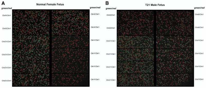
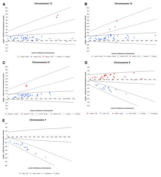
Study design: Twenty-four amniocentesis and 16 chorionic villus samples were used for microfluidic digital PCR analysis. Three thousand and sixty PCR reactions were performed for each of the target chromosomes (X, Y, 13, 18, and 21), and the number of single molecule amplifications was compared to a reference. The difference between target and reference chromosome counts was used to determine the ploidy of each of the target chromosomes.
Results: Digital PCR accurately identified all cases of fetal trisomy (3 cases of trisomy 21, 3 cases of trisomy 18, and 2 cases of triosmy 13) in the 40 specimens analyzed. The remaining specimens were determined to have normal ploidy for the chromosomes tested.
Conclusion: Microfluidic digital PCR allows detection of fetal chromosomal aneuploidy utilizing uncultured amniocytes and chorionic villus tissue in less than 6 hours.
Figures
Similar articles
-
Use of a DNA method, QF-PCR, in the prenatal diagnosis of fetal aneuploidies.J Obstet Gynaecol Can. 2011 Sep;33(9):955-960. doi: 10.1016/S1701-2163(16)35022-8. J Obstet Gynaecol Can. 2011. PMID: 21923994 Review.
-
Chip-Based Digital PCR Approach Provides A Sensitive and Cost-Effective Single-Day Screening Tool for Common Fetal Aneuploidies-A Proof of Concept Study.Int J Mol Sci. 2019 Nov 4;20(21):5486. doi: 10.3390/ijms20215486. Int J Mol Sci. 2019. PMID: 31690017 Free PMC article.
-
Rapid prenatal diagnosis of aneuploidy for chromosomes 21, 18, 13, and X by quantitative fluorescence polymerase chain reaction.Fetal Diagn Ther. 2006;21(4):326-31. doi: 10.1159/000092459. Fetal Diagn Ther. 2006. PMID: 16757905
-
Rapid detection of chromosomal aneuploidies in uncultured amniocytes by multiplex ligation-dependent probe amplification (MLPA).Prenat Diagn. 2005 Nov;25(11):1032-9. doi: 10.1002/pd.1247. Prenat Diagn. 2005. PMID: 16231311
-
Genomic medicine in prenatal diagnosis.Clin Obstet Gynecol. 2008 Mar;51(1):62-73. doi: 10.1097/GRF.0b013e3181616509. Clin Obstet Gynecol. 2008. PMID: 18303500 Review.
Cited by
-
Non-invasive prenatal diagnosis using cell-free fetal nucleic acids in maternal plasma: Progress overview beyond predictive and personalized diagnosis.EPMA J. 2011 Jun;2(2):163-71. doi: 10.1007/s13167-011-0085-y. Epub 2011 May 17. EPMA J. 2011. PMID: 23199146 Free PMC article.
-
The Influence of Maternal Cell Contamination on Fetal Aneuploidy Detection Using Chip-Based Digital PCR Testing.Diagnostics (Basel). 2021 Sep 3;11(9):1607. doi: 10.3390/diagnostics11091607. Diagnostics (Basel). 2021. PMID: 34573950 Free PMC article.
-
Microfluidics in reproductive biology: applying lab-on-a-chip technologies to assisted reproduction.Mol Hum Reprod. 2017 Apr 1;23(4):211-212. doi: 10.1093/molehr/gax020. Mol Hum Reprod. 2017. PMID: 28369546 Free PMC article. No abstract available.
-
Universal noninvasive detection of solid organ transplant rejection.Proc Natl Acad Sci U S A. 2011 Apr 12;108(15):6229-34. doi: 10.1073/pnas.1013924108. Epub 2011 Mar 28. Proc Natl Acad Sci U S A. 2011. PMID: 21444804 Free PMC article.
-
Next-generation sequencing of 35 RHD variants in 16 253 serologically D- pregnant women in the Finnish population.Blood Adv. 2020 Oct 27;4(20):4994-5001. doi: 10.1182/bloodadvances.2020001569. Blood Adv. 2020. PMID: 33057632 Free PMC article.
References
-
- Cunningham F, Hauth J, Leveno K, Gilstrap L, Bloom S, Wenstrom K. Williams obstetrics. New York: McGraw-Hill Professional; 2002. p. 942.
-
- ACOG Practice Bulletin No. 88, December 2007. Invasive prenatal testing for aneuploidy. Obstet Gynecol. 2007;110:1459–67. - PubMed
-
- Hulten MA, Dhanjal S, Pertl B. Rapid and simple prenatal diagnosis of common chromosome disorders: advantages and disadvantages of the molecular methods FISH and QF-PCR. Reproduction. 2003;126:279–97. - PubMed
-
- Dudarewicz L, Holzgreve W, Jeziorowska A, Jakubowski L, Zimmermann B. Molecular methods for rapid detection of aneuploidy. J Appl Genet. 2005;46:207–15. - PubMed
-
- Shaffer LG, Bui TH. Molecular cytogenetic and rapid aneuploidy detection methods in prenatal diagnosis. Am J Med Genet C Semin Med Genet. 2007;145C:87–98. - PubMed
Publication types
MeSH terms
Grants and funding
LinkOut - more resources
Full Text Sources
Other Literature Sources
Medical
