. 2009 Jun 12;344(9):1072-8.
doi: 10.1016/j.carres.2009.03.026. Epub 2009 Mar 27.

Real-time NMR monitoring of intermediates and labile products of the bifunctional enzyme UDP-apiose/UDP-xylose synthase

Affiliations

Real-time NMR monitoring of intermediates and labile products of the bifunctional enzyme UDP-apiose/UDP-xylose synthase

Paul Guyett et al. Carbohydr Res. .

Abstract

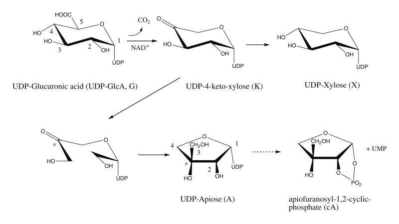
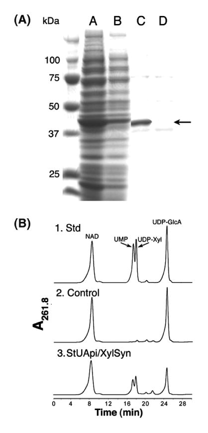
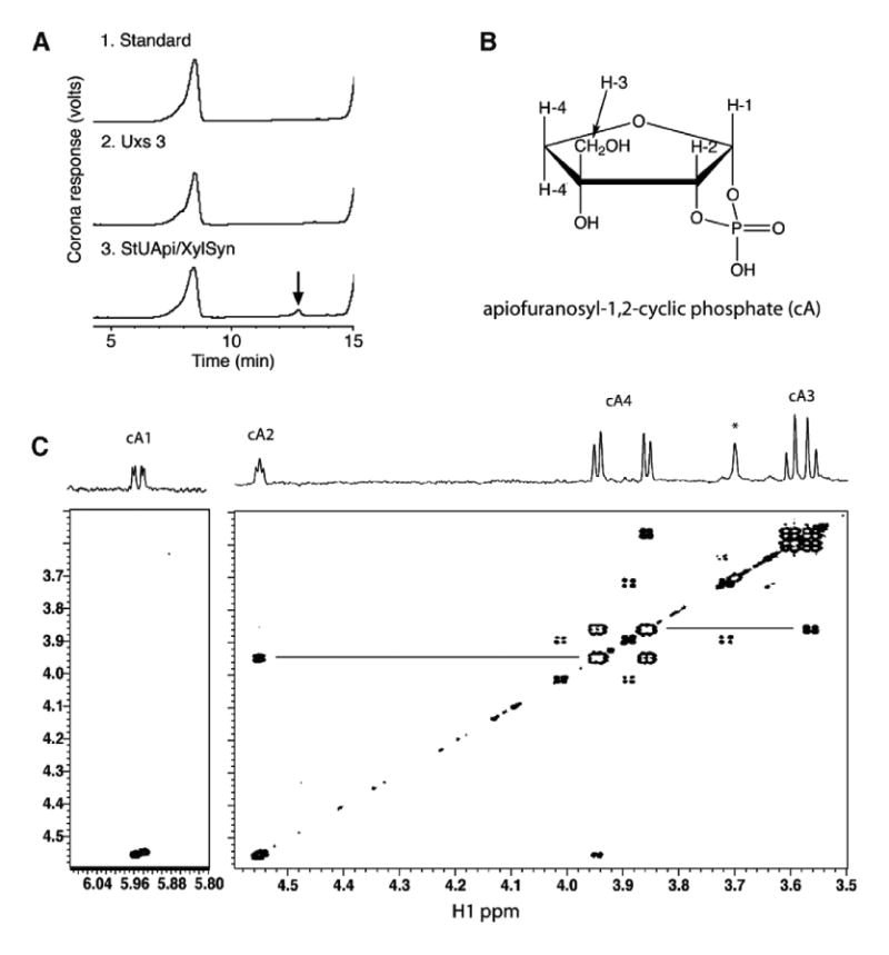
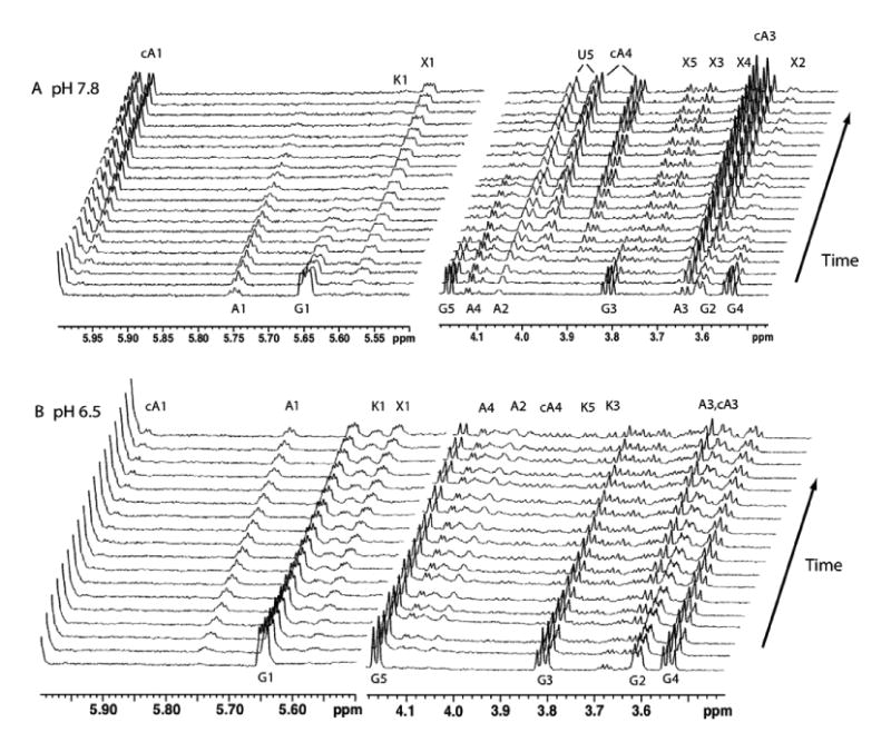
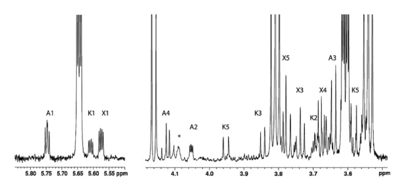
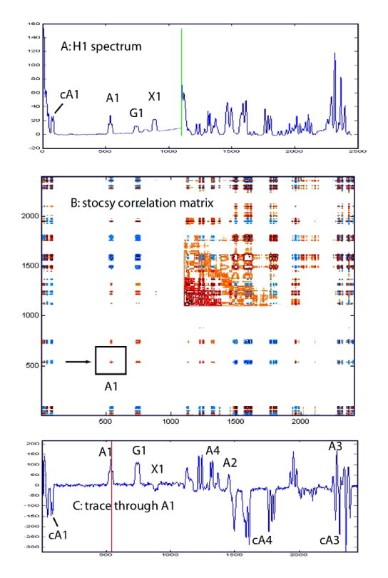
The conversion of UDP-alpha-d-glucuronic acid to UDP-alpha-d-xylose and UDP-alpha-d-apiose by a bifunctional potato enzyme UDP-apiose/UDP-xylose synthase was studied using real-time nuclear magnetic resonance (NMR) spectroscopy. UDP-alpha-d-glucuronic acid is converted via the intermediate uridine 5'-beta-l-threo-pentapyranosyl-4''-ulose diphosphate to UDP-alpha-d-apiose and simultaneously to UDP-alpha-d-xylose. The UDP-alpha-d-apiose that is formed is unstable and is converted to alpha-d-apio-furanosyl-1,2-cyclic phosphate and UMP. High-resolution real-time NMR spectroscopy is a powerful tool for the direct and quantitative characterization of previously undetected transient and labile components formed during a complex enzyme-catalyzed reaction.


Figures

Figure 1
Figure 1
Proposed mechanism for the conversion of UDP-GlcA to UDP-xylose and UDP-apiose catalyzed by UAXS. The UDP-4-keto-xylose structure is depicted in the keto form, but it is most likely in equilibrium with the hydrated gem-diol form. The numbering schemes of the UDP-GlcA and UDP-apiose are independent. Carbon 4 of UDP-GlcA (marked with an asterisk) becomes carbon 3 of UDP-apiose. Note that the ring protons have been left off for clarity.
Figure 2
Figure 2
Protein expression of UAXS and analysis of enzymatic reaction using HPLC. Panel A: SDS–PAGE of soluble protein isolated from E. coli cell expressing recombinant UAXS (lane A) or control empty vector (lane B). Column fractionation of UAXS (lane C) compared to an equivalent fraction from cell containing the empty vector (lane D). The band indicated by an arrow is the over-expressed recombinant UAXS. Panel B: (1) HPLC analysis of standard NAD+, UMP, UDP-xylose, and UDPGlcA, (2) reaction products when UDP-GlcA and NAD+ are reacted with protein from control empty vector expression, and (3) reaction products when UDP-GlcA and NAD+ are reacted with the recombinant UAXS.
Figure 3
Figure 3
Identification of apiofuranosyl-1,2-cyclic phosphate (cA). Panel A: Ion-exchange HPLC chromatogram of products formed when UDP-GlcA and NAD+ were reacted with the recombinant UAXS. A peak (marked by arrow) was detected when the reaction mixture contained UAXS but was absent in the control reactions. Panel B: Chemical structure: in this numbering scheme the exocyclic methylene protons are labeled as H3, and ring methylene protons are labeled as H4. (C) COSY NMR spectrum: Four-bond couplings between H2 and H4, and H3 and H4, are indicated by horizontal lines. The signals are labeled as cA1, etc. to conform to subsequent figures.
Figure 4
Figure 4
Real-time 1H NMR analysis of the products formed when recombinant UAXS is reacted at pH 7.8 in 80% D2O-20% H2O–phosphate buffer at 35 °C with UDP-GlcA and NAD+ (Panel A, upper set). Regions of the 800-MHz proton spectra are plotted at 25-min intervals starting at the bottom, from 15 min after the addition of UAXS. Signals are labeled as originating from UDP-GlcA (G), UDP-apiose (A), apiofuranosyl-1,2-cyclic phosphate (cA), UDP-xylose (X), UDP-4-keto-xylose (K), and the ribose of UMP (U). Panel B, lower set: UAXS activity with UDP-GlcA and NAD+ when reactions were carried out at pH 6.5 in 80% D2O–20% H2O–phosphate buffer at 35 °C Spectra are plotted at 48-min intervals starting at the bottom (15 min). Signals are labeled as in Panel A.
Figure 5
Figure 5
Regions of a 900-MHz 1H NMR spectrum of the reaction mixture after 2 h at pH 6.5 in 90% H2O buffer at 33 °C. Signals are labeled as UDP-apiose (A), UDP-xylose (X), and UDP-4-keto-xylose (K). No detectable amounts of apiofuranosyl-1,2-cyclic phosphate have been formed at this stage. The signal labeled A4 shows the expected AB quartet pattern of methylene protons, and the signal labeled A2 shows scalar coupling to A1 and long-range coupling to 31P. The broad signal at 4.09 ppm indicated with an asterisk is correlated to UDP-apiose and is tentatively assigned as a ribose H5 proton.
Figure 6
Figure 6
Covariance matrix representation (STOCSY) of the spectra shown in Figure 4A corresponding to the time interval from 90 to 490 min. During this period, the UDP-apiose converts to apiofuranosyl-1,2-cyclic phosphate. Top spectrum A shows the corresponding regions from a 1D spectrum of the reaction mixture. Bottom spectrum C shows the peaks that are correlated to UDP-apiose resonance A1, indicated by box in panel C. See text for further discussion.

References

    1. Harborne JB, Williams CA. Nat Prod Rep. 2001;18:310–333. - PubMed
    1. Su YF, Koike K, Guo D, Satou T, Liu JS, Zheng JH, Nikaido T. Tetrahedron. 2001;57:6721–6726.
    1. El-Sayed NH, Wojcinska M, Drost-Karbowska K, Matlawska I, Williams J, Mabry TJ. Phytochemistry. 2002;60:835–838. - PubMed
    1. Siciliano T, De Tommasi N, Morelli I, Braca A. J Agric Food Chem. 2004;52:6510–6515. - PubMed
    1. Darvill AG, O'Neill MA, Albersheim P. Plant Physiol. 1978;62:418–422. - PMC - PubMed

Publication types

MeSH terms

LinkOut - more resources