

Mechanism of shortened bones in mucopolysaccharidosis VII
- PMID: 19375967
- PMCID: PMC2775472
- DOI: 10.1016/j.ymgme.2009.03.005
Mechanism of shortened bones in mucopolysaccharidosis VII
Abstract
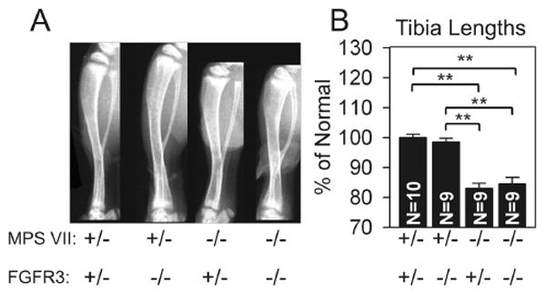
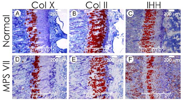
Mucopolysaccharidosis VII (MPS VII) is a lysosomal storage disease in which deficiency in beta-glucuronidase results in glycosaminoglycan (GAG) accumulation in and around cells, causing shortened long bones through mechanisms that remain largely unclear. We demonstrate here that MPS VII mice accumulate massive amounts of the GAG chondroitin-4-sulfate (C4S) in their growth plates, the cartilaginous region near the ends of long bones responsible for growth. MPS VII mice also have only 60% of the normal number of chondrocytes in the growth plate and 55% of normal chondrocyte proliferation at 3weeks of age. We hypothesized that this reduction in proliferation was due to C4S-mediated overactivation of fibroblast growth factor receptor 3 (FGFR3). However, MPS VII mice that were FGFR3-deficient still had shortened bones, suggesting that FGFR3 is not required for the bone defect. Further study revealed that MPS VII growth plates had reduced tyrosine phosphorylation of STAT3, a pro-proliferative transcription factor. This was accompanied by a decrease in expression of leukemia inhibitory factor (LIF) and other interleukin 6 family cytokines, and a reduction in phosphorylated tyrosine kinase 2 (TYK2), Janus kinase 1 (JAK1), and JAK2, known activators of STAT3 phosphorylation. Intriguingly, loss of function mutations in LIF and its receptor leads to shortened bones. This suggests that accumulation of C4S in the growth plate leads to reduced expression of LIF and reduced STAT3 tyrosine phosphorylation, which results in reduced chondrocyte proliferation and ultimately shortened bones.
Figures


Similar articles
-
Neonatal retroviral vector-mediated hepatic gene therapy reduces bone, joint, and cartilage disease in mucopolysaccharidosis VII mice and dogs.Mol Genet Metab. 2004 May;82(1):4-19. doi: 10.1016/j.ymgme.2004.01.015. Mol Genet Metab. 2004. PMID: 15110316
-
Delayed hypertrophic differentiation of epiphyseal chondrocytes contributes to failed secondary ossification in mucopolysaccharidosis VII dogs.Mol Genet Metab. 2015 Nov;116(3):195-203. doi: 10.1016/j.ymgme.2015.09.008. Epub 2015 Sep 26. Mol Genet Metab. 2015. PMID: 26422116 Free PMC article.
-
Progression of vertebral bone disease in mucopolysaccharidosis VII dogs from birth to skeletal maturity.Mol Genet Metab. 2021 Aug;133(4):378-385. doi: 10.1016/j.ymgme.2021.06.005. Epub 2021 Jun 15. Mol Genet Metab. 2021. PMID: 34154922 Free PMC article.
-
Murine mucopolysaccharidosis type VII: the impact of therapies on the clinical course and pathology in a murine model of lysosomal storage disease.J Inherit Metab Dis. 1998 Aug;21(5):575-86. doi: 10.1023/a:1005423222927. J Inherit Metab Dis. 1998. PMID: 9728337 Review.
-
Gene therapy for murine mucopolysaccharidosis type VII.Neuromuscul Disord. 1997 Jul;7(5):352-60. doi: 10.1016/s0960-8966(97)00061-8. Neuromuscul Disord. 1997. PMID: 9267850 Review.
Cited by
-
MPSI Manifestations and Treatment Outcome: Skeletal Focus.Int J Mol Sci. 2022 Sep 22;23(19):11168. doi: 10.3390/ijms231911168. Int J Mol Sci. 2022. PMID: 36232472 Free PMC article. Review.
-
Lysosomal and network alterations in human mucopolysaccharidosis type VII iPSC-derived neurons.Sci Rep. 2018 Nov 9;8(1):16644. doi: 10.1038/s41598-018-34523-3. Sci Rep. 2018. PMID: 30413728 Free PMC article.
-
Biomarkers for prediction of skeletal disease progression in mucopolysaccharidosis type I.JIMD Rep. 2020 Dec 8;58(1):89-99. doi: 10.1002/jmd2.12190. eCollection 2021 Mar. JIMD Rep. 2020. PMID: 33728251 Free PMC article.
-
Long circulating enzyme replacement therapy rescues bone pathology in mucopolysaccharidosis VII murine model.Mol Genet Metab. 2012 Sep;107(1-2):161-72. doi: 10.1016/j.ymgme.2012.07.002. Epub 2012 Jul 14. Mol Genet Metab. 2012. PMID: 22902520 Free PMC article.
-
Anti-TNF-alpha therapy enhances the effects of enzyme replacement therapy in rats with mucopolysaccharidosis type VI.PLoS One. 2011;6(8):e22447. doi: 10.1371/journal.pone.0022447. Epub 2011 Aug 22. PLoS One. 2011. PMID: 21887218 Free PMC article.
References
-
- Neufeld EF, Meunzer J. The mucopolysaccharidoses. In: Scriver CR, Beaudet AL, Sly WS, Valle D, editors. Metabolic and Molecular Basis of Inherited Disease. McGraw-Hill; New York: 2001. pp. 3421–3452.
-
- Pastores GM, Arn P, Beck M, Clarke JTR, Guffon N, Kaplan P, Muenzer J, Norato DYJ, Shapiro E, Thomas J, Viskochil D, Wraith JE. The MPS I registry: design, methodology, and early findings of a global disease registry for monitoring patients with Mucopolysaccharidosis Type I. Mol Genet Metab. 2007;91:37–47. - PubMed
-
- Mango RL, Xu L, Sands MS, Vogler C, Seiler G, Schwartz T, Haskins ME, Ponder KP. Neonatal retroviral vector-mediated hepatic gene therapy reduces bone, joint, and cartilage disease in mucopolysaccharidosis VII mice and dogs. Mol Gen Metab. 2004;82:4–19. - PubMed
Publication types
MeSH terms
Substances
Grants and funding
LinkOut - more resources
Full Text Sources
Other Literature Sources
Medical
Molecular Biology Databases
Research Materials
Miscellaneous
