. 2009 Apr 16;62(1):72-83.
doi: 10.1016/j.neuron.2009.02.020.

Activation of the p75 neurotrophin receptor through conformational rearrangement of disulphide-linked receptor dimers

Affiliations

Activation of the p75 neurotrophin receptor through conformational rearrangement of disulphide-linked receptor dimers

Marçal Vilar et al. Neuron. .

Abstract

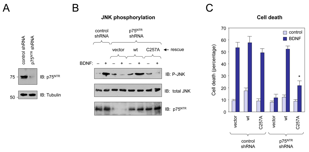
Ligand-mediated dimerization has emerged as a universal mechanism of growth factor receptor activation. Neurotrophins interact with dimers of the p75 neurotrophin receptor (p75(NTR)), but the mechanism of receptor activation has remained elusive. Here, we show that p75(NTR) forms disulphide-linked dimers independently of neurotrophin binding through the highly conserved Cys(257) in its transmembrane domain. Mutation of Cys(257) abolished neurotrophin-dependent receptor activity but did not affect downstream signaling by the p75(NTR)/NgR/Lingo-1 complex in response to MAG, indicating the existence of distinct, ligand-specific activation mechanisms for p75(NTR). FRET experiments revealed a close association of p75(NTR) intracellular domains that was transiently disrupted by conformational changes induced upon NGF binding. Although mutation of Cys(257) did not alter the oligomeric state of p75(NTR), the mutant receptor was no longer able to propagate conformational changes to the cytoplasmic domain upon ligand binding. We propose that neurotrophins activate p75(NTR) by a mechanism involving rearrangement of disulphide-linked receptor subunits.


Figures

Figure 1
Figure 1. Transmembrane Cys257 mediates formation of disulphide-linked p75NTR dimers
(A) Cell surface expression of disulphide-linked p75NTR dimers (dim) and monomers (mon) in transfected COS-7 cells visualized by Neutravidin probing of p75NTR immunoprecipitates under non-reducing (−DTT) and reducing (+DTT) conditions. In non-reducing conditions, p75NTR dimers run somewhat higher, and monomers lower, than their predicted molecular weights. (B) Cell surface expression of disulphide-linked p75NTR dimers (dim) and monomers (mon) in COS-7 cells transfected with different amounts of wild type p75NTR. Immunoprecipitates were electrophoresed under non-reducing conditions and p75NTR was visualized by Neutravidin probing of p75NTR immunoprecipitates. Results are expressed as mean dimer/monomer ratio. (C) Alignment of p75NTR transmembrane domain sequences from vertebrate and invertebrate species. (D) Cell surface expression of endogenous disulphide-linked p75NTR dimers (dim) and monomers (mon) in PC12 cells, RN22 Schwannoma cells and SCG sympathetic neurons visualized by Neutravidin probing of p75NTR immunoprecipitates. Control, untransfected COS cells. (E) Expression of endogenous disulphide-linked p75NTR dimers (dim) and monomers (mon) in extracts of newborn rat cerebellum (cblm), cortex (ctx) and hippocampus (hc) visualized by immunoblotting of p75NTR immunoprecipitates under reducing and non-reducing conditions. The control lane represents a mock immunoprecipitate of the cblm extract. (F) Binding of 125I-NGF to wild type and C257A p75NTR analyzed by chemical crosslinking. Samples were run under reducing conditions. For each construct, binding counts were normalized to levels of expression as assessed by immunoblotting (IB). Results are expressed as mean ± SD of three independent determinations. (G) γ-secretase-dependent intramembrane cleavage of C257A p75NTR following stimulation with PMA in transfected COS cells. The proteasome inhibitor epoxomycin was used to prevent degradation of CTF and ICD fragments. Mutant and wild type (wt) p75NTR molecules were recovered by immunoprecipitation and visualized by immunoblotting. The γ-secretase inhibitor DAPT blocked the generation of p75NTR intracellular domain (ICD). CTF, carboxy terminal fragment.
Figure 2
Figure 2. Cys257 is essential for recruitment of NRIF and TRAF6 to p75NTR in response to NGF
(A) Binding of NRIF to wild type and mutant p75NTR in transfected HEK293 cells assayed by immunoprecipitation (IP) and immunoblotting (IB). (B) Binding of TRAF6 to wild type and mutant p75NTR in transfected HEK293 cells. (C)Binding of NRIF to wild type and mutant p75NTR in transfected HEK293 cells. Prior to gel electrophoresis, the pull-down sample was splitted in two equal parts, one was boiled in sample buffer, the other was treated with 1M DTT prior to boiling. The migration of p75NTR dimers and monomers is indicated. (D) Binding of TRAF6 to wild type and mutant p75NTR in transfected HEK293 cells analyzed as in panel (C). (E) Binding of NRIF to wild type and mutant p75NTR in rat SCG neurons. NRIF only interacts with p75NTR disulphide-linked dimers, not monomers.
Figure 3
Figure 3. Cys257 is essential for p75NTR signaling to NF-κB, caspase-3 and cell death in response to NGF
(A) NF-κB activity in transfected M23 fibroblasts in the presence and absence of NGF. (B) Activation of caspase-3 visualized with a cleavage-specific antibody in HEK293 cells transfected with p75NTR constructs in response to NGF. Reprobing controls for p75NTR and GAPDH are shown. Independent experiments confirmed comparable expression of transfected p75NTR constructs. (C) Cell death assay in HEK293 cells transfected with p75NTR constructs in response to NGF. Results are expressed as mean ± SD of three independent experiments, each performed in duplicate.
Figure 4
Figure 4. Cys257 is essential for p75NTR signaling in SCG neurons
(A) Downregulation of p75NTR expression in rat SCG neurons following transfection of p75NTR shRNAs. (B) Assay of JNK phosphorylation in SCG neurons in response to BDNF. SCG neurons were transfected with the indicated shRNA constructs. In rescue experiments, wild type p75NTR or C257A p75NTR constructs insensitive to p75NTR shRNAs were also introduced by DNA transfection. JNK phosphorylation was assessed by immunoblotting of total cell lysates. (C) Cell death in SCG neurons transfected with p75NTR shRNAs in response to BDNF. Rescue experiments with wild type and C257A p75NTR constructs were performed as above. *, p<0.05 vs. wild type, n=3.
Figure 5
Figure 5. Cys257 is not required for downstream signaling by the p75NTR/NgR/Lingo-1 complex in response to MAG
(A) Binding of RhoGDI to wild type and C257A p75NTR in COS-7 cells co-transfected with NgR and Lingo-1 and stimulated with MAG-Fc. (B) RhoA activity in COS-7 cells transfected with wild type and mutant p75NTR in the presence of NgR and Lingo-1 after stimulation with MAG-Fc for 30 min. Results are expressed as mean ± SD relative to wild type without MAG treatment. *, p<0.05 vs. control, n=3.
Figure 6
Figure 6. Cell surface p75NTR dimerization is mediated by covalent and non-covalent interactions between transmembrane domains
(A) Wild type and C257A p75NTR dimers analyzed by cell surface chemical crosslinking under reducing conditions. Cell lysates were immunoprecipitated with anti-p75NTR antibodies; immunoblot was probed with anti-HA antibodies. (B) Alignment of p75NTR transmembrane domains highlighting the AxxxG self-association motif and Gly266. (C) ToxCAT assay of self-association of wild type and mutant p75NTR transmembrane domains. Wild type and mutant transmembrane domains from glycophorin A (GpATM) were used as positive and negative controls, respectively. (D) Wild type and mutant p75NTR dimers analyzed by cell surface chemical crosslinking as above.
Figure 7
Figure 7. Analysis of conformational changes in p75NTR intracellular domains by anisotropy microscopy
(A–D) Steady-state anisotropy in transfected cells. Examples of areas used for anisotropy measurements are boxed and shown as high magnification insets. Monomeric EGFP (EGFP1, high anisotropy, low FRET) and a concatenated EGFP trimer (EGFP3, low anisotropy, high FRET) were used as controls. The calibration bar of the look-up table is shown below. (E) Steady-state anisotropy of wild type and C257A p75NTR-EGFP in COS-7 cells. The anisotropy value of EGFP1 was arbitrarily set to zero and used as baseline for the histogram. Bars show average ± SD (n=8–11 cells for EGFP and 22–30 cells for p75NTR). (F) Representative examples of anisotropy traces after addition of NGF or medium in cells expressing wild type or C257A p75NTR-EGFP. A peak in anisotropy was observed after NGF addition in all cells expressing wild type p75NTR that were examined (n=30) regardless of their initial baseline anisotropy level. (G) Anisotropy change after addition of NGF or medium (control). The difference in anisotropy before and after addition of NGF (i.e. peak minus baseline value) or medium was calculated for wild type and C257A p75NTR-EGFP. Results are expressed as average ± SD (n=15 to 17 cells examined). *, p<0.0001 vs. C257A.
Figure 8
Figure 8. The “snail-tong” mechanism of p75NTR activation in response to neurotrophins
Hypothetical schematic of p75NTR in the cell membrane before and after neurotrophin binding (adapted from Gong et al. (2008) and Liepinsh et al. (1997)). The approximate position of Cys257 is indicated. Arrows denote the postulated “snail-tong” movement of p75NTR subunits initiated by ligand binding: closing onto the neurotrophin dimer in the outside, opening in the inside.

Comment in

References

    1. Ashkenazi A, Dixit VM. Apoptosis control by death and decoy receptors. Curr Opin Cell Biol. 1999;11:255–260. - PubMed
    1. Barker PA. p75NTR is positively promiscuous: novel partners and new insights. Neuron. 2004;42:529–533. - PubMed
    1. Bibel M, Barde YA. Neurotrophins: key regulators of cell fate and cell shape in the vertebrate nervous system. Genes Dev. 2000;14:2919–2937. - PubMed
    1. Bothwell M. Evolution of the neurotrophin signaling system in invertebrates. Brain Behav Evol. 2006;68:124–132. - PubMed
    1. Bronfman FC, Fainzilber M. Multi-tasking by the p75 neurotrophin receptor: sortilin things out? EMBO Rep. 2004;5:867–871. - PMC - PubMed

Publication types

MeSH terms