. 2009 Jul;1788(7):1458-65.
doi: 10.1016/j.bbamem.2009.03.022. Epub 2009 Apr 17.

Protein 4.1R links E-cadherin/beta-catenin complex to the cytoskeleton through its direct interaction with beta-catenin and modulates adherens junction integrity

Affiliations

Protein 4.1R links E-cadherin/beta-catenin complex to the cytoskeleton through its direct interaction with beta-catenin and modulates adherens junction integrity

Shaomin Yang et al. Biochim Biophys Acta. 2009 Jul.

Abstract

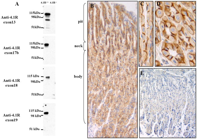
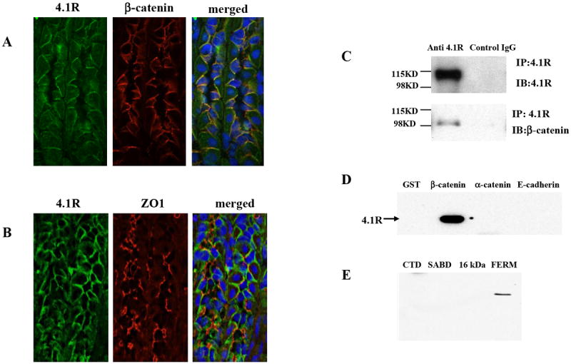
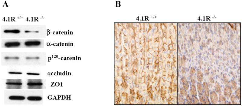
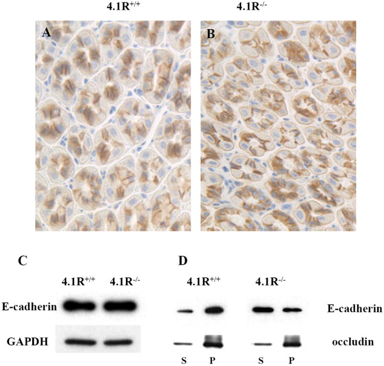
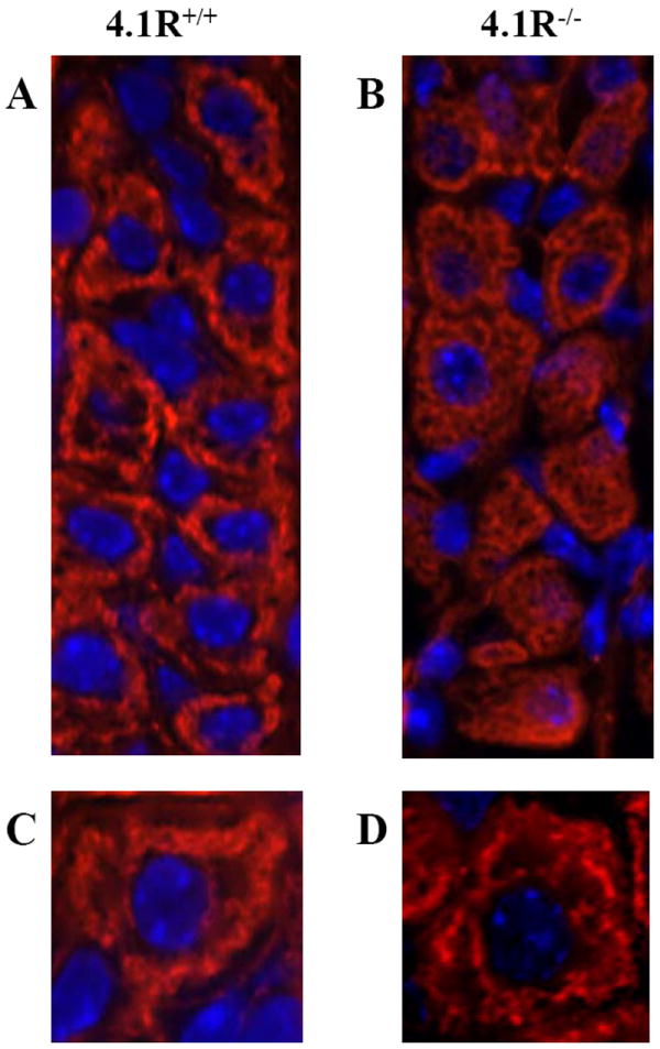
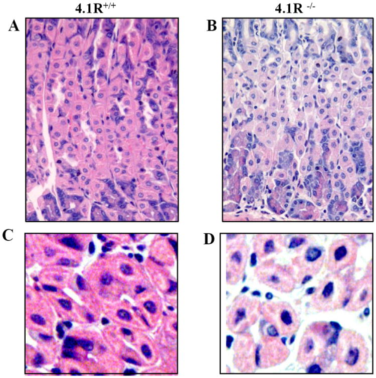
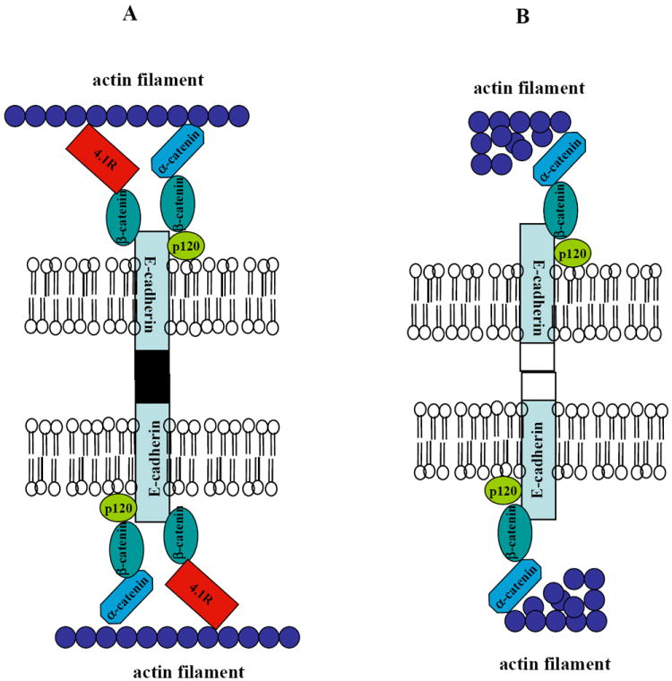
Protein 4.1R (4.1R) is the prototypical member of the protein 4.1 superfamily comprising of the protein 4.1 family (4.1R, 4.1B, 4.1G and 4.1N) and ERM family (ezrin, radixin and meosin). These proteins in general serve as adaptors between the membrane and the cytoskeleton. Here we show that 4.1R expressed in the gastric epithelial cells associates with adherens junction protein beta-catenin. Biochemical examination of 4.1R-deficient stomach epithelia revealed a selective reduction of beta-catenin which is accompanied by a weaker linkage of E-cadherin to the cytoskeleton. In addition, organization of actin cytoskeleton was altered in 4.1R-deficient cells. Moreover, histological examination revealed that cell-cell contacts are impaired and gastric glands are disorganized in 4.1R null stomach epithelia. These results demonstrate an important and previously unidentified role of 4.1R in linking the cadherin/catenin complex to the cytoskeleton through its direct interaction with beta-catenin and in regulating the integrity of adherens junction.


Figures

Fig 1
Fig 1. Expression and Localization of Protein 4.1R on Gastric Epithelia
(A) Western blotting analysis of 4.1R from gastric mucosa. Total cell lysates from wild-type and 4.1R-/-mice mucosa were probed with antibodies against 4.1R exon13, exon17b, exon18 and exon19, while anti-exon 13 antibody detected an ~80 kDa and a ~100 kDa band, the other three antibodies only detected the ~100 kDa band. (B-E) Immunolocalization of 4.1R on gastric epithelia. 4.1R is expressed in all layers of gastric epithelia, with strong staining in the middle part of the gastric glands, where parietal cells predominate (B, 20x). Part of the neck region was observed under higher magnification (C, longitudinal section; D, transverse section, 40x). Note positive signal mainly on the bilateral membrane of the cells. No staining was observed in corresponding sections from 4.1R-/- mice (E, 20x).
Fig 2
Fig 2. Association of 4.1R with β-catenin
(A) Co-localization of 4.1R with β-catenin. Frozen sections from wild-type stomach were co-stained with goat anti-4.1R exon13 antibody (green) and rabbit anti-β-catenin antibody (red). Merged image shows that 4.1R co-localizes with β-catenin on the bilateral membrane. (B) 4.1R does not co-localize with ZO-1. Frozen sections from wild-type stomach were co-stained with goat anti-4.1R exon13 antibody (green) and rabbit anti ZO-1 antibody (red). Merged image shows distinct localization of 4.1R and ZO-1. (C) Co-immunoprecipation of β-catenin with 4.1R. 4.1R was immunoprecipated from from 4.1R+/+ gastric mucosa lysate using anti-4.1R or pre-immune IgG. 4.1R or β-catenin in the immunoprecipate was detected using anti-4.1R antibody (upper panel) or anti-β-catenin antibody (lower panel) respectively. Note that β-catenin was brought down with 4.1R. (D) Binding of 4.1R to β-catenin. Recombinant His-tagged 4.1R was incubated with GST, GST-tagged β-catenin, GST-tagged α-catenin or GST-tagged cytoplasmic domain of E-cadherin. Binding was assayed by pull down assay, using anti-His antibody for detection. Note that 4.1R only bound to β-catenin. (E) Binding of 4.1R domains to β-catenin. Recombinant His-tagged 4.1R domains (FERM domain; SABD: spectrin-actin binding domain; CTD: C-terminal domain) were incubated with GST-tagged β-catenin. Binding was assayed as above, using anti-His antibody for detection. Note that only FERM bound to β-catenin.
Fig 3
Fig 3. Selective reduction of β-catenin in 4.1R-/- gastric epithelia
(A) Western blotting analysis. Expression of β-catenin, α-catenin, p120 catenin, ZO-1 and occudin in gastric mucosa was examined by Western blotting analysis. Note the selective reduction of β-catenin in 4.1R-/-gastric mucosa. (B) Immunohistochemistry. β-catenin was stained in paraffin-embedded wild type and 4.1R-/- stomach tissue sections. Note the staining of β-catenin on the bilateral membrane of wild-type gastric epithelial cells (left panel) and the reduction of it in the 4.1R-/- stomach.
Fig 4
Fig 4. Weakened attachment of E-cadherin to cytoskeleton in 4.1R-/- gastric epithelia
(A and B) Immunostaining of E-cadherin in wild-type and 4.1R-/- gastric epithelia. E-cadherin is located at the bilateral membranes of both wild-type (A) and 4.1R-/-(B) gastric epithelial cells. (C) Western blotting analysis of E-cadherin from gastric mucosa. Total cell lysates from mucosa of wild-type and 4.1R-/- mice were probed with anti-E-cadherin antibody. No difference was observed between wild type and 4.1R-/- mice. GAPDH was used as loading control. (D) Increased detergent extractability of E-cadherin from 4.1R-/- gastric mucosa. Transmembane proteins were extracted from wild type and 4.1R-/- gastric mucosa with buffer containing 0.5% Triton X-100. Western blotting analysis was performed using anti-E-cadherin or anti-occudin antibodies. Note retention of the bulk of E-cadherin in the pellet from wild-type samples, the greater part is recovered in the supernatant from 4.1R-/- samples. No difference was observed in the retention of tight junction protein occudin between wild type and 4.1R-/-.
Fig 5
Fig 5. Impaired F-actin cytoskeleton organization in 4.1R-/- gastric epithelial cells
F-actin filaments were stained with rhodamine-phalloidin. In wild-type cells (A and C), actin filaments are distributed in a sharp, well-defined thread-like pattern around nucleus (displaying the intracellular secretory canalicular system), in the 4.1R-/- cells (B and D) it is uniformly distributed throughout the cytoplasm. Blue is DAPI staining for the nucleus. Magnifications: x40 (A, B); x100 (C, D).
Fig 6
Fig 6. Disorganization of glands and impaired cell-cell adhesion of 4.1R-/- Gastric Epithelia
(A and B): the images are of H&E-stained sections of wild-type and 4.1R-/- gastric epithelia under 40x magnification. Note the disorganized glands of 4.1R-/- gastric epithelium. (C and D): enlarged images from A and B reveal tightly packed cells and pyramidal or polygonal parietal cells in the wild-type. Note the irregular outline of gastric glands, rounded cells with short lateral membranes and loose cell-cell contact in 4.1R -/- gastric epithelia.
Fig 7
Fig 7. Working model for role of 4.1R in gastric epithelial cells
(A) In wild type cell, E-cadherin/catenin complex is well attached to the actin cytoskeleton by both α-catenin and 4.1R. This ensures the adhesive activity of E-cadherin. (B) In 4.1R null cells, β-catenin is reduced and the remaining β-catenin is linked the actin cytoskeleton only by α-catenin. This resulted in weakened attachment of E-cadherin to the skeleton and the impaired cell-cell adhesion. In addition, actin filament is also disorganized.

Similar articles

Cited by

References

    1. Conboy J, Kan YW, Shohet SB, Mohandas N. Molecular cloning of protein 4.1, a major structural element of the human erythrocyte membrane skeleton. Proc Natl Acad Sci U S A. 1986;83:9512–6. - PMC - PubMed
    1. Parra M, Gascard P, Walensky LD, Gimm JA, Blackshaw S, Chan N, Takakuwa Y, Berger T, Lee G, Chasis JA, Snyder SH, Mohandas N, Conboy JG. Molecular and functional characterization of protein 4.1B, a novel member of the protein 4.1 family with high level, focal expression in brain. J Biol Chem. 2000;275:3247–55. - PubMed
    1. Parra M, Gascard P, Walensky LD, Snyder SH, Mohandas N, Conboy JG. Cloning and characterization of 4.1G (EPB41L2), a new member of the skeletal protein 4.1 (EPB41) gene family. Genomics. 1998;49:298–306. - PubMed
    1. Walensky LD, Blackshaw S, Liao D, Watkins CC, Weier HU, Parra M, Huganir RL, Conboy JG, Mohandas N, Snyder SH. A novel neuron-enriched homolog of the erythrocyte membrane cytoskeletal protein 4.1. J Neurosci. 1999;19:6457–67. - PMC - PubMed
    1. Gould KL, Bretscher A, Esch FS, Hunter T. cDNA cloning and sequencing of the protein-tyrosine kinase substrate, ezrin, reveals homology to band 4.1. Embo J. 1989;8:4133–42. - PMC - PubMed

Publication types

LinkOut - more resources