. 2009 Aug 1;442(1-2):55-62.
doi: 10.1016/j.gene.2009.04.003. Epub 2009 Apr 17.

Activation of the brain-specific neurogranin gene in murine T-cell lymphomas by proviral insertional mutagenesis

Affiliations

Activation of the brain-specific neurogranin gene in murine T-cell lymphomas by proviral insertional mutagenesis

Anne Ahlmann Nielsen et al. Gene. .

Abstract

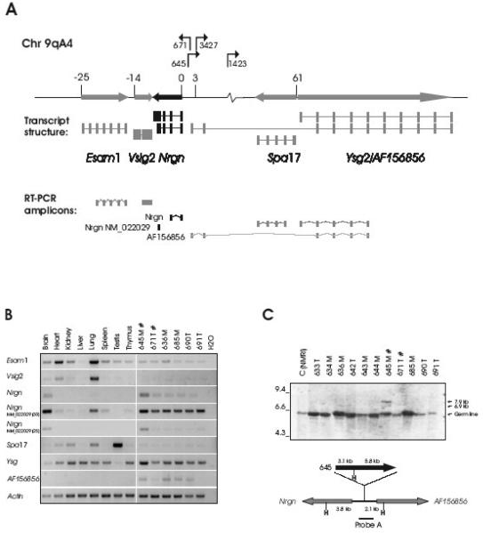
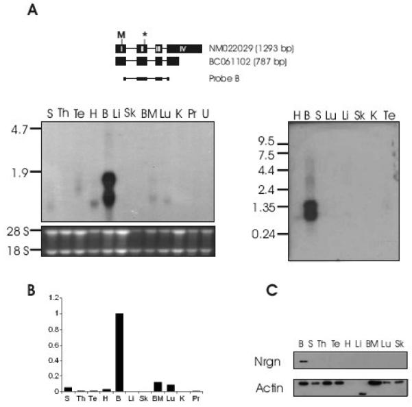
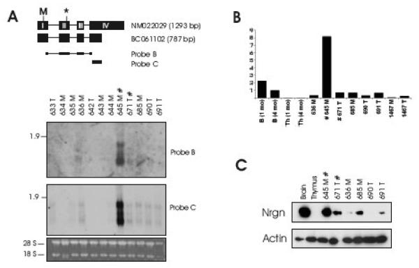
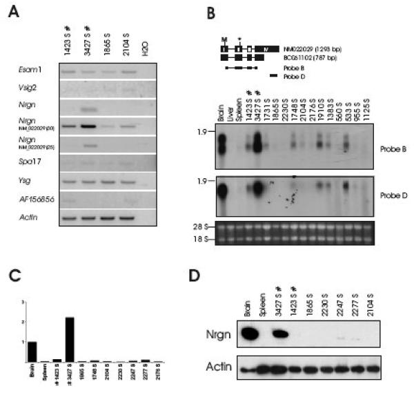
Neurogranin (Nrgn) is a highly expressed brain-specific protein, which sequesters calmodulin at low Ca(2+)-levels. We report here on retroviral activation of the Nrgn gene in tumors induced by the T-cell lymphomagenic SL3-3 murine leukemia virus. We have performed a systematic expression analysis of Nrgn in various mouse tissues and SL3-3 induced T-cell tumors. This demonstrated that insertional activation of Nrgn increased RNA and protein expression levels to that observed in brain. Furthermore, elevated Nrgn expression was also observed in some T-cell tumors with no detected provirus integrations into this genomic region. The presented data demonstrate that Nrgn can be produced at high levels outside the brain, and suggest a novel oncogenic role in T-cell lymphomas in mice.


Figures

Fig. 1
Fig. 1. Proviral integrations in the Esam1/Vsig2/Nrgn/Ysg2/Spa17-locus activate Nrgn expression in T-cell tumors
A. The proviral positions and transcriptional orientations in the locus are shown with arrows. The relative position of the transcription start sites of the genes are given in kb and a schematic mRNA structure is depicted with exons as bars. Both a long (NM_022029) and a short (BC061102) transcript form of Nrgn is shown in black. The RT-PCR amplicons are drawn below. B. RT-PCR on RNA from a panel of tissues from non-infected mice (lanes 1-8) as well as six independent mesenteric (‘M’) and thymic (‘T’) tumors induced by SL3-3(turbo) (lanes 9-14). Tumors with integration into the Nrgn locus are indicated with ‘#’. For the ‘NM_022029’ amplicon PCR amplification products with 25 and 30 cycles are shown. C. Southern blotting on HindIII-digested tumor DNA using Probe A (top panel). Positions of the germ line band as well as the sizes of the expected rearranged bands (6.9 kb and 7.9 kb) are indicated with arrows. The position of Probe A across the integration sites between Nrgn and AF156856 is depicted schematically together with HindIII sites (‘H’), and the distances in kb between HindIII positions and the integration site (bottom panel). For clarity, only the clonal provirus in tumor 645 is depicted. ‘T’ and ‘M’ designates a thymic or mesenteric lymph node tumor, respectively.
Fig. 2
Fig. 2. Expression of Nrgn is highest in brain
A. Northern blotting using Probe B positioned on the long and short Nrgn mRNA as shown schematically (top panel). The extent of CDS is indicated by start (‘M’) and termination (‘*’) codon. Northern blotting was performed on a mRNA Multiple Northern Blot (Clontech) (right panel) as well as on a membrane containing total RNA from various organs from BALB/c mice (left panel). H, heart; B, brain; S, spleen; Lu, lung; Li, liver; Sk, skeletal muscle; K, kidney; Te, testis; Th, thymus; BM, bone marrow; Pr, prostate; U, uterus. B. Relative Nrgn levels as measured by quantitative real-time PCR. C. Nrgn Western blotting on protein isolated from a panel of mouse organs.
Fig. 3
Fig. 3. Neurogranin expression in SL3-3(turbo) MLV-induced T-cell lymphomas equals that of brain tissue
Northern blot analysis (A), Quantitative real-time PCR (B) and Western blotting (C) on tumors from SL3-3(turbo)-infected mice. Northern blotting was done using the indicated probes. In (B) brain and thymus from 1 month and 4 month old non-infected mice were included for comparison. In (C) brain and thymus from 1 month old non-infected mice were included. Legend is as in Fig. 1.
Fig. 4
Fig. 4. Nrgn is expressed in SL3-3 wt MLV-induced T-cell lymphomas
(A) RT-PCR analysis using gene-specific primers as in Fig. 1 is shown. ‘S’ designates splenic tumor. Northern blotting with the indicated probes (B), Quantitative real-time PCR (C) and Western blotting (D) was performed as described for Fig. 3.

Similar articles

Cited by

References

    1. Akagi K, Suzuki T, Stephens RM, Jenkins NA, Copeland NG. RTCGD: retroviral tagged cancer gene database. Nucl. Acids. Res. 2004;32:D523–527. - PMC - PubMed
    1. Baudier J, Deloulme JC, Van Dorsselaer A, Black D, Matthes HW. Purification and characterization of a brain-specific protein kinase C substrate, neurogranin (p17). Identification of a consensus amino acid sequence between neurogranin and neuromodulin (GAP43) that corresponds to the protein kinase C phosphorylation site and the calmodulin-binding domain. J Biol Chem. 1991;266:229–37. - PubMed
    1. Chakravarthy B, Morley P, Whitfield J. Ca2+-calmodulin and protein kinase Cs: a hypothetical synthesis of their conflicting convergences on shared substrate domains. Trends Neurosci. 1999;22:12–6. - PubMed
    1. Chiriva-Internati M, Wang Z, Salati E, Timmins P, Lim SH. Tumor vaccine for ovarian carcinoma targeting sperm protein 17. Cancer. 2002;94:2447–53. - PubMed
    1. Chretien I, Marcuz A, Courtet M, Katevuo K, Vainio O, Heath JK, White SJ, Du Pasquier L. CTX, a Xenopus thymocyte receptor, defines a molecular family conserved throughout vertebrates. Eur J Immunol. 1998;28:4094–104. - PubMed

Publication types

LinkOut - more resources