

White matter abnormalities revealed by diffusion tensor imaging in non-demented and demented HIV+ patients
- PMID: 19376246
- PMCID: PMC4494862
- DOI: 10.1016/j.neuroimage.2009.04.030
White matter abnormalities revealed by diffusion tensor imaging in non-demented and demented HIV+ patients
Abstract
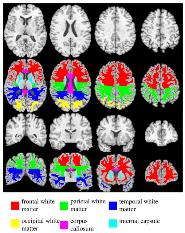
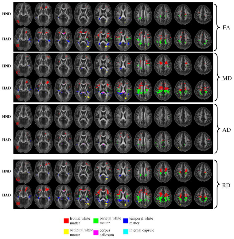
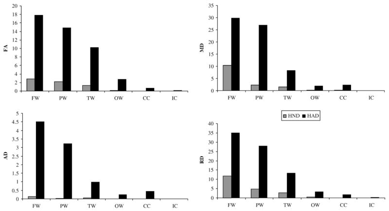
HIV associated dementia (HAD) is the most advanced stage of central nervous system disease caused by HIV infection. Previous studies have demonstrated that patients with HAD exhibit greater cerebral and basal ganglia atrophy than non-demented HIV+ (HND) patients. However, the extent to which white matter is affected in HAD patients compared to HND patients remains elusive. This study is designed to address the potential white matter abnormalities through the utilization of diffusion tensor imaging (DTI) in both HND and HAD patients. DTI and T1-weighted images were acquired from 18 healthy controls, 21 HND and 8 HAD patients. T1 image-based registration was performed to 1) parcellate the whole brain white matter into major white matter regions, including frontal, parietal, temporal and occipital white matter, corpus callosum and internal capsule for statistical comparisons of the mean DTI values, and 2) warp all DTI parametric images towards the common template space for voxel-based analysis. The statistical comparisons were performed with four DTI parameters including fractional anisotropy (FA), mean (MD), axial (AD), and radial (RD) diffusivities. With Whitney U tests on the mean DTI values, both HND and HAD demonstrated significant differences from the healthy control in multiple white matter regions. In addition, HAD patients exhibited significantly elevated MD and RD in the parietal white matter when compared to HND patients. In the voxel-based analysis, widespread abnormal regions were identified for both HND and HAD patients, although a much larger abnormal volume was observed in HAD patients for all four DTI parameters. Furthermore, both region of interest (ROI) based and voxel-based analyses revealed that RD was affected to a much greater extent than AD by HIV infection, which may suggest that demyelination is the prominent disease progression in white matter.
Figures


References
-
- AIDSTaskForce. Nomenclature and research case definitions for neurologic manifestations of human immunodeficiency virus-type 1 (HIV-1) infection. Report of a Working Group of the American Academy of Neurology AIDS Task Force. Neurology. 1991;41:778–785. - PubMed
-
- Albright AV, Soldan SS, Gonzalez-Scarano F. Pathogenesis of human immunodeficiency virus-induced neurological disease. J Neurovirol. 2003;9:222–227. - PubMed
-
- An SF, Scaravilli F. Early HIV-1 infection of the central nervous system. Arch Anat Cytol Pathol. 1997;45:94–105. - PubMed
-
- Aylward EH, Brettschneider PD, McArthur JC, Harris GJ, Schlaepfer TE, Henderer JD, Barta PE, Tien AY, Pearlson GD. Magnetic resonance imaging measurement of gray matter volume reductions in HIV dementia. Am J Psychiatry. 1995;152:987–994. - PubMed
-
- Aylward EH, Henderer JD, McArthur JC, Brettschneider PD, Harris GJ, Barta PE, Pearlson GD. Reduced basal ganglia volume in HIV-1-associated dementia: results from quantitative neuroimaging. Neurology. 1993;43:2099–2104. - PubMed
MeSH terms
Grants and funding
LinkOut - more resources
Full Text Sources
Medical
Miscellaneous
