Review
. 2009 May;28(3):155-77.
doi: 10.1016/j.preteyeres.2009.04.003. Epub 2009 Apr 17.

Neural regulation of lacrimal gland secretory processes: relevance in dry eye diseases

Affiliations
Review

Neural regulation of lacrimal gland secretory processes: relevance in dry eye diseases

Darlene A Dartt. Prog Retin Eye Res. 2009 May.

Abstract

The lacrimal gland is the major contributor to the aqueous layer of the tear film which consists of water, electrolytes and proteins. The amount and composition of this layer is critical for the health, maintenance, and protection of the cells of the cornea and conjunctiva (the ocular surface). Small changes in the concentration of tear electrolytes have been correlated with dry eye syndrome. While the mechanisms of secretion of water, electrolytes and proteins from the lacrimal gland differ, all three are under tight neural control. This allows for a rapid response to meet the needs of the cells of the ocular surface in response to environmental conditions. The neural response consists of the activation of the afferent sensory nerves in the cornea and conjunctiva to stimulate efferent parasympathetic and sympathetic nerves that innervate the lacrimal gland. Neurotransmitters are released from the stimulated parasympathetic and sympathetic nerves that cause secretion of water, electrolytes, and proteins from the lacrimal gland and onto the ocular surface. This review focuses on the neural regulation of lacrimal gland secretion under normal and dry eye conditions.


Figures
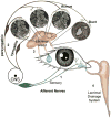
Figure 1
Figure 1. Lacrimal Gland Functional Unit
Schematic of the components of the lacrimal gland functional unit that includes: (1) Activation of afferent sensory nerves from the cornea and conjunctiva that project through the central nervous system (CNS) to stimulate (2) Efferent parasympathetic and sympathetic nerves that innervate the lacrimal gland acinar and ductal cells and induce (3) Secretion of lacrimal gland fluid containing proteins, electrolytes, and water through the duct system onto the ocular surface that (4) drains into the lacrimal gland into the lacrimal drainage system. Modified from Rocha et al The Ocular Surface 6:162-174, 2008.
Figure 2
Figure 2. Schematic of the Structure of the Lacrimal Gland
This schematic shows the pyramidally shaped acinar cells linked at the luminal side by tight junctions that generate polarized secretion of proteins, electrolytes and water. Parasympathetic and sympathetic nerves release neurotransmitters to activate their receptors on the basolateral membranes. Stimulation of these receptors induces a series of second messenger components that activate: (1) ion channels and pumps differentially located on the basolateral and apical membranes neurotransmitters to cause electrolyte and water secretion and (2) protein secretion by exocytosis. Ach-acetylcholine, M3AChR-type 3 muscarinc acetylcholine receptor, VIP-vasoactive intestinal peptide, VIPACR1-VIP receptor type 1, α1D-AR- α adrenergic receptor type 1D, EGF- epidermal growth factor, EGFR-epidermal growth factor receptor
Figure 3
Figure 3. Immunofluorescence micrograph of lacrimal gland myoepithelial cells
Myoepithelial cells of rat lacrimal gland stained with an antibody to α smooth muscle actin. Myoepithelial cells are stellate-shaped cells that surround the basal aspect of acinar and ductal cells that are below the plane of focus.
Figure 4
Figure 4. Schematic of the functional types of sensory neurons innervating the ocular surface
Four types of neurons are described with their differential location, basal activity, stimulated activity, and receptor type. Reprinted with permission from Belmonte et al The Ocular Surface 2:248, 2004.
Figure 5
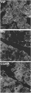
Figure 5. Immunofluorescence micrographs of the distribution of parasympathetic, sympathetic, and sensory nerves in the lacrimal gland
Mouse lacrimal gland incubated with antibodies against various neurotransmitters. Anti-VIP demonstrates location of parasympathetic nerves surrounding most acini. Anti-tyrosine hydroxylase (TH) indicates sympathetic nerves that surround a few acinar. Anticalcitonin gene-related peptide (CGRP) shows sensory nerves with a limited distribution.
Figure 6
Figure 6. Overview of neural signaling pathways in the lacrimal gland
Stimulation of parasympathetic and sympathetic nerves releases their neurotransmitters to activate their receptors on the basolateral membranes. Stimulation of these receptors induces a series of stimulatory and inhibitory second messenger components that are specific for each neurotransmitter and induce protein secretion by exocytosis. Ach-acetylcholine, M3AChR- type 3 muscarinc acetylcholine receptor, VIP-vasoactive intestinal peptide, VIPACR1- VIP receptor type 1, α1D-AR- α adrenergic receptor type 1D, EGF- epidermal growth factor, EGFR-epidermal growth factor receptor, PLC-phospholipase C, PLD-phospholipase D, PKC- protein kinase C, MAPK-p44/p42 mitogen-activated protein kinase, NO-nitric oxide.
Figure 7
Figure 7. Schematic representation of parasympathetic signaling pathway
Acetycholine (Ach) is released from parasympathic nerves and binds to M3 muscarinic receptor (M3AChR). This initiates a signaling cascade that involves activation of phospholipase Cβ (PLCβ) and protein kinase C (PKC)α, ε, and δ, and the formation of diacylglycerol (DAG) and 1,4,5-inositol trisphosphate (IP3). Calcium is released from the endoplasmic reticulum (ER) leading to protein secretion. The M3AChR also activates non-receptor tyrosine kinases Pyk2 and Src to activate Ras, Raf, mitogen activated protein kinase kinase (MEK) and p44/p42 mitogen-activated protein kinase (MAPK) that attenuates protein secretion.
Figure 8
Figure 8. Schematic representation of molecular mechanism of capacitative calcium entry
Agonist activation of a plasma membrane receptor results in formation of inositol-1,4,5-trisphosphate (IP3), which activates the IP3 receptor causing discharge of store Ca2+ from a subcompartment of the endoplasmic reticulum. Within this subcompartment, Ca2+ binds reversibly to an EF hand motif in Stim1; depletion of Ca2+ results in Stim1 without Ca2+ bound, which causes Stim1 to redistribute within the endoplasmic reticulum to areas near Orai1 within the plasma membrane. Stim1 then activates Ca2+-selective Orai channels; the mechanism whereby this activation is accomplished in unknown. TRPC are additional mechanism for Ca2+ entry as they are non-selective cation channels that allow Ca2+ entry. Ag- agonist, G- guanine nucleotide-binding protein, PLC-phospholipase C, TRPC- transient receptor potential channel. Reprinted with permission from Putney Cell Calcium 42(2):103-110, 2007.
Figure 9
Figure 9. Schematic Representation of VIP signaling pathway
VIP released from parasympathetic nerves binds to its receptors (VIPACR1 and II) and activates adenylyl cyclase (AC) to produce increase cellular levels of cAMP. cAMP activates protein kinase A (PKA) that stimulates protein secretion. Gαs- stimulatory guanine nucleotide-binding protein α subunit.
Figure 10
Figure 10. Schematic representation of the different components of A-kinase anchoring proteins (AKAPs)
There are numerous types of AKAPs that each bind a distinct set of signaling molecules in addition to protein kinase A (PKA). The Gravin AKAP binds phosphodiesterase. The muscle (m)AKAP binds phosphodiesterase and EPAC1 (exchange protein activated by cAMP) and extracellular regulated kinase (ERK, a mitogen-activated protein kinase). AKAP79 binds adenylyl cyclase. R-regulatory subunit of PKA, C- catalytic subunit of PKA. PDE- cAMP-dependent phosphodiesterase. Reprinted with persmission from Beene and Scott, Curr Opin Cell Biol 19(2), 192-198. 2007.
Figure 11
Figure 11. Schematic of α1D-adrenergic signaling pathway
Norepinephrine is released from sympathetic nerves to bind to the α1D-adrenergic receptors (α1D-ARs) that activates protein kinase C (PKC)ε and endothelial nitric oxide synthase (eNOS) to stimulate lacrimal gland protein secretion. α1D-ARs also activate PKCα and –δ to attenuate protein secretion. In addition, matrix metalloproteinases (MMP) are activated by these recptors to transactivate the EGF receptor activating the Ras signaling cascade that also attenuates protein secretion. Gαq- guanine nucleotide-binding protein; NO – nitric oxide; GC – guanylate cyclase; Raf – mitogen activated protein kinase kinase kinase; MEK - mitogen activated protein kinase kinase; MAPK - mitogen activated protein kinase.
Figure 12
Figure 12. Schematic of the Mechanism of Interaction of Ca2+/protein kinase C and cAMP-dependent signaling pathways
The muscarinic, cholinergic Ca2+/protein kinase C-dependent signaling pathway is illustrated here as an example of the EGF and α1D-adrenergic signaling pathways. VIP stimulated increase in cAMP is illustrated as an example of mechanisms that increase cAMP. VIP released from parasympathetic nerves binds to its receptors (VIPACR1 and 2) and activates adenylyl cyclase (AC) to increase cellular levels of cAMP. cAMP activates protein kinase A (PKA) that inhibitis activation of p44/p42 mitogen-activated protein kinase (MAPK) by cholinergic agonists. Cholinergic agonists, through increasing the intracellular [Ca2+] and activate protein kinase C (PKC), activate the non-receptor tyrosine kinases Pyk2 and Src to activate MAPK. Gαs-stimulatory guanine nucleotide-binding protein, PDE-cAMP-dependent phosphodiesterase, Ras, Raf-mitogen-activated protein kinase kinase kinase, MEK-mitogen-activated protein kinase kinase.
Figure 13
Figure 13. Schematic of working model for mechanism of basal and stimulated exocytosis for protein secretion
(A) Resting lacrimal gland acini maintain mature secretory vesicles (mSVs) enriched in Rab3D beneath dense apical filament network. The mSVs utilize cytoplasmic dynein for their maturation and to maintain their subapical loacalization. Other recruitable SVs (rSVs) enriched in VAMP2 are present in the cytosol. (B) Upon agonist stimulation there is an immediate thinning of apical actin filaments concomitant with the loss of Rab3D from primed individual SVs in clusters accompanying assembly of actin and non-muscle myosin II filaments around these clusters. Contractile forces generated by the acto-myosin system results in compound fusion of individual SVs and movement of these fusion intermediates toward the apical plasma membrane (APM). Ultimately, they reach the APM where they are able to gain access to appropriate SNARE proteins and complete the fusion step. At the same time, rSVs in the cytosol move along the microtubules (MTs) with the aid of cytoplasmic dynein to the APM, where they fuse and release their contents through formation of a SNARE pair with rSV VAMP2 and the APM syntaxin 3. Reprinted with permission from Wu et al Experimental Eye Research 83:84-96, 2006.
Figure 14
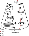
Figure 14. Schematic of transcytosis mechanism of protein secretion
Secretion of dimeric IgA (dIgA) illustrates the transcytosis secretory mechanism. Polymeric IgA receptor (pIgR) trafficks dIgA leading to secretion of secretory IgA and secretory component (SC). PigR and SC can be secreted by exocytosis via the process described in Figure 13 and shown with black arrows. These compounds can also be secreted by transcytosis by the process illustrated in red arrows. ER-endoplasmic reticulum, BE-basolateral endosomes, TGN-transgolgi network, AE- apical endosomes, SV- secretory vesicles. Reprinted with permission from Evans et al Am J Physiology Cell Physiol 294:C662-C674, 2008.
Figure 15
Figure 15. Mechanism of electrolyte and water secretion by lacrimal gland acinar cells
NKA, Na+,K+-ATPase, AE, anion (Cl-/HCO3-) exchanger, NHE, Na+/H+ exchanger, NKCC- Na+-K+-2Cl- cotransporter. Reprinted with permission from Selvam et al Am J Cell Physiol 293:C1412-C1419, 2007.
Figure 16
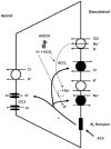
Figure 16. Model for secretion of electrolytes and water by lacrimal gland duct cells
Parasympathetic agonists increase the intracellular [Ca2+] that stimulates the Na+/H+ exchanger (NHE) followed by activation of the anion (Cl-/HCO3-) exchanger (AE) on the basolateral membrane that drives Na+ and Cl- into the duct cells. The Na+ and Cl-is dependent upon the generation of H+ and HCO3- generated by intracellular carbonic anhydrase (CA). The elevated Na+ can be exchanged for K+ through the basolateral Na+,K+-ATPase, which increases cellular K+ concentration. The increase in intracellular K+ and Cl- drives these ions out of the cell into the lumen via specific K+ and Cl-channels (IKCa1 and CIC3, respectively) and a K+/Cl- co-transporter. An increase in intracellular [Ca2+] can also stimulate IKCa1. Reprinted with permission from Toth-Molnar et al Invest Ophthalmol Vis Sci 48(8);3746-3755,2007.
Figure 17
Figure 17. Schematic Representation of the neural regulation of lacrimal gland secretion in the diseased state
Proinflammatory cytokines block the release of neurotransmitters from the afferent sensory nerves and the efferent parasympathetic and sympathetic nerves preventing their stimulation of lacrimal gland fluid secretion resulting in an inadequate secretion leading to aqueous deficiency dry eye. Modified from Dartt et al The Ocular Surface 2:76-91, 2004.

References

    1. Acosta MC, Peral A, Luna C, Pintor J, Belmonte C, Gallar J. Tear secretion induced by selective stimulation of corneal and conjunctival sensory nerve fibers. Invest Ophthalmol Vis Sci. 2004;45(7):2333–2336. - PubMed
    1. Acosta MC, Tan ME, Belmonte C, Gallar J. Sensations evoked by selective mechanical, chemical, and thermal stimulation of the conjunctiva and cornea. Invest Ophthalmol Vis Sci. 2001;42(9):2063–2067. - PubMed
    1. Allansmith MR, Kajiyama G, Abelson MB, Simon MA. Plasma cell content of main and accessory lacrimal glands and conjunctiva. Am J Ophthalmol. 1976;82(6):819–826. - PubMed
    1. Andersson SV, Hamm-Alvarez SF, Gierow JP. Integrin adhesion in regulation of lacrimal gland acinar cell secretion. Exp Eye Res. 2006;83(3):543–553. - PubMed
    1. Arslan G, Fredholm BB. Stimulatory and inhibitory effects of adenosine A(2A) receptors on nerve growth factor-induced phosphorylation of extracellular regulated kinases 1/2 in PC12 cells. Neurosci Lett. 2000;292(3):183–186. - PubMed

Substances

LinkOut - more resources