

Effects of age on optical coherence tomography measurements of healthy retinal nerve fiber layer, macula, and optic nerve head
- PMID: 19376593
- PMCID: PMC2747246
- DOI: 10.1016/j.ophtha.2009.01.004
Effects of age on optical coherence tomography measurements of healthy retinal nerve fiber layer, macula, and optic nerve head
Abstract
Purpose: To determine the effects of age on global and sectoral peripapillary retinal nerve fiber layer (RNFL), macular thicknesses, and optic nerve head (ONH) parameters in healthy subjects using optical coherence tomography (OCT).
Design: Retrospective, cross-sectional observational study.
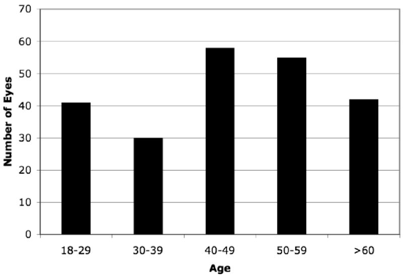
Participants: A total of 226 eyes from 124 healthy subjects were included.
Methods: Healthy subjects were scanned using the Fast RNFL, Fast Macula, and Fast ONH scan patterns on a Stratus OCT (Carl Zeiss Meditec, Dublin, CA). All global and sectoral RNFL and macular parameters and global ONH parameters were modeled in terms of age using linear mixed effects models. Normalized slopes were also calculated by dividing the slopes by the mean value of the OCT parameter for interparameter comparison.
Main outcome measures: Slope of each OCT parameter across age.
Results: All global and sectoral RNFL thickness parameters statistically significantly decreased with increasing age, except for the temporal quadrant and clock hours 8 to 10, which were not statistically different from a slope of zero. Highest absolute slopes were in the inferior and superior quadrant RNFL and clock hour 1 (superior nasal). Normalized slopes showed a similar rate in all sectors except for the temporal clock hours (8-10). All macular thickness parameters statistically significantly decreased with increasing age, except for the central fovea sector, which had a slight positive slope that was not statistically significant. The nasal outer sector had the greatest absolute slope. Normalized macular slope in the outer ring was similar to the normalized slopes in the RNFL. Normalized inner ring had shallower slope than the outer ring with a similar rate in all quadrants. Disc area remained nearly constant across the ages, but cup area increased and rim area decreased with age, both of which were statistically significant.
Conclusions: Global and regional changes caused by the effects of age on RNFL, macula, and ONH OCT measurements should be considered when assessing eyes over time.
Financial disclosure(s): Proprietary or commercial disclosure may be found after the references.
Conflict of interest statement
Conflict of Interest: Dr. Wollstein received research funding from Carl Zeiss Meditec and Optovue. Drs. Wollstein, Ishikawa and Schuman receive royalties for intellectual property licensed by the University of Pittsburgh to Biopigen. Drs. Schuman and Fujimoto receive royalties for intellectual property licensed by Massachusetts Institute of Technology to Carl Zeiss Meditec. Dr. Fujimoto is a scientific advisor and has stock options with Optovue.
References
-
- Villain MA, Greenfield DS. Peripapillary nerve fiber layer thickness measurement reproducibility using optical coherence tomography. Ophthalmic Surg Lasers Imaging. 2003;34:33–7. - PubMed
-
- Budenz DL, Chang RT, Huang X, et al. Reproducibility of retinal nerve fiber thickness measurements using the Stratus OCT in normal and glaucomatous eyes. Invest Ophthalmol Vis Sci. 2005;46:2440–3. - PubMed
-
- Blumenthal EZ, Williams JM, Weinreb RN, et al. Reproducibility of nerve fiber layer thickness measurements by use of optical coherence tomography. Ophthalmology. 2000;107:2278–82. - PubMed
Publication types
MeSH terms
Grants and funding
LinkOut - more resources
Full Text Sources
Medical
