. 2009 Jun;46(3):359-69.
doi: 10.1016/j.cyto.2009.03.010. Epub 2009 Apr 18.

Tumor necrosis factor (TNF)-alpha-induced IL-8 expression in gastric epithelial cells: role of reactive oxygen species and AP endonuclease-1/redox factor (Ref)-1

Affiliations

Tumor necrosis factor (TNF)-alpha-induced IL-8 expression in gastric epithelial cells: role of reactive oxygen species and AP endonuclease-1/redox factor (Ref)-1

Ann M O'Hara et al. Cytokine. 2009 Jun.

Abstract

TNF-alpha contributes to oxidative stress via induction of reactive oxygen species (ROS) and pro-inflammatory cytokines. The molecular basis of this is not well understood but it is partly mediated through the inducible expression of IL-8. As redox factor-1 (Ref-1), is an important mediator of redox-regulated gene expression we investigated whether ROS and Ref-1 modulate TNF-alpha-induced IL-8 expression in human gastric epithelial cells. We found that TNF-alpha treatment of AGS cells enhanced nuclear expression of Ref-1 and potently induced IL-8 expression. Overexpression of Ref-1 enhanced IL-8 gene transcription at baseline and after TNF-alpha treatment whereas Ref-1 suppression and antioxidant treatment inhibited TNF-alpha-stimulated IL-8 expression. TNF-alpha-mediated enhancement of other pro-inflammatory chemokines like MIP-3 alpha and Gro-alpha was also regulated by Ref-1. Although TNF-alpha increased DNA binding activity of Ref-1-regulated transcription factors, AP-1 and NF-kappaB, to the IL-8 promoter, promoter activity was mainly mediated by NF-kappaB binding. Silencing of Ref-1 in AGS cells inhibited basal and TNF-alpha-induced AP-1 and NF-kappaB DNA binding activity, but not their nuclear accumulation. Collectively, we provide the first mechanistic evidence of Ref-1 involvement in TNF-alpha-mediated, redox-sensitive induction of IL-8 and other chemokines in human gastric mucosa. This has implications for understanding the pathogenesis of gastrointestinal inflammatory disorders.


Figures
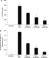
Fig. 1
Fig. 1
NAC inhibits TNF-α-induced IL-8 protein (A) and mRNA (B) production in AGS cells. AGS cells were pre-treated with 0 mM, 10 mM, 20 mM, or 40 mM NAC for 30 min prior to treatment with 20 ng/ml TNF-α or left untreated (controls). After 3 h the supernatants were assayed for IL-8 protein by ELISA (A). TNF-α stimulated the secretion of IL-8 protein by AGS cells, and pre-treatment with 20 mM and 40 mM NAC significantly suppressed TNF-α-induced IL-8 protein (*P < 0.05 versus cells that were treated with TNF-α only). (B) IL-8 mRNA expression was assayed by real-time RT-PCR 1 h post-treatment, and the graph depicts the levels of IL-8 mRNA (normalized to 18S rRNA) detected. IL-8 mRNA expression was increased by TNF-α treatment when compared to untreated cells, and pre-treatment with NAC at all concentrations tested (10, 20, or 40 mM NAC) significantly decreased TNF-α-induced IL-8 mRNA (* P < 0.05 versus cells that were treated with TNF-α only). Results represent the mean ± SEM (n = 3).
Fig. 2
Fig. 2
(A) Effect of Ref-1 suppression on TNF-α-induced IL-8 expression in AGS cells. AGS cells transfected with 50 nM Ref-1 siRNA or mock-transfected cells were treated 48 h post-transfection with 20 ng/ml TNF-α or left untreated. Our previous studies demonstrated that IL-8 expression was no different in AGS cells that were transfected with scrambled siRNA versus mock-transfected cells [32]. IL-8 mRNA expression levels were assayed by real-time RT-PCR 1 h post-treatment. The graph depicts the percent IL-8 mRNA expression (normalized to 18S rRNA) of TNF-α-stimulated cells transfected with Ref-1 siRNA compared to TNF-α-stimulated cells transfected with Lipofectamine 2000 alone. TNF-α-induced IL-8 mRNA expression was significantly inhibited (*P < 0.05) in AGS cells transfected with Ref-1 siRNA compared to cells transfected with Lipofectamine 2000 alone. (B) Forty-eight hours post-transfection, cells were stimulated with TNF-α for 3 h and the supernatants were assayed for IL-8 by ELISA. Results are expressed as pg IL-8/mg total protein concentration. In cells expressing normal levels of Ref-1, TNF-α stimulated a significant increase in IL-8 protein. In cells transfected with Ref-1 siRNA, TNF-α-induced IL-8 protein was significantly inhibited (*P < 0.05 when compared to TNF-α-stimulated mock-transfected cells). All data represent the mean ± SEM of three separate experiments.
Fig. 3
Fig. 3
siRNA-mediated silencing of Ref-1 inhibits TNF-α-induced AP-1 and NF-κB DNA binding activity but does not block nuclear accumulation of these transcription factors. AGS cells transfected with 50 nM Ref-1 siRNA (+) or non-transfected (−) cells were treated with 20 ng/ml TNF-α 48 h post-transfection. Untreated cells served as controls. Nuclear extracts were used to bind to radiolabeled AP-1 or NF-κB probes. (A) A representative EMSA autoradiogram showing AP-1 activation in non-transfected or Ref-1 siRNA-transfected AGS cells treated for 1 h with TNF-α. The arrow indicates the activated AP-1 complex. (B) A representative EMSA autoradiogram showing NF-κB activation in non-transfected or Ref-1 siRNA-transfected AGS cells treated with TNF-α for 2 h. The arrow indicates the activated NF-κB complex. The specificity of the shifted AP-1 and NF-κB complexes were analyzed using a 50-fold excess of unlabeled specific competitor or unlabeled non-specific competitor as indicated. (C) Non-transfected control cells (lanes 1 and 4), and cells transfected with 50 nM control siRNA (lanes 2 and 5) or Ref-1 siRNA (lanes 3 and 6) were treated with 20 ng/ml TNF-α or left untreated 48 h post-transfection for 1 or 2 h. After harvesting, nuclear extracts were analyzed by SDS–PAGE immunoblotting for NF-κB p50, p65, c-Jun, c-Fos. Ref-1 levels were assessed in nuclear and total cellular extracts. The data shown are representative of four separate experiments.
Fig. 4
Fig. 4
Schematic representation of the luciferase-linked human IL-8 promoter constructs used in this study. The plasmids contain sequentially deleted 5′ flanking regions of the human IL-8 gene. Locations of transcription factor binding sites are indicated. The AP-1 binding site is located between –126 and –120 nucleotides, the NF-IL-6 binding site is between –94 and –81 nucleotides, and the NF-κB binding site is between –80 and –70 nucleotides. A consensus TATA box is located between –20 and –13 nucleotides.
Fig. 5
Fig. 5
Effect of 5′ deletions and site-directed mutations in the human IL-8 promoter sequence on TNF-α-inducible IL-8 luciferase activity in cells with normal and over-expressed levels of Ref-1. AGS cells transfected with 5′ deletions of the hIL-8 Luc promoter were treated with 20 ng/ml TNF-α for 3 h. Untreated AGS cells served as controls. IL-8 luciferase activity normalized to protein, was expressed as fold induction for each construct relative to untreated cells (A). In cells bearing the −1498/+44 or the −99/+44 hIL-8/Luc constructs, TNF-α significantly stimulated IL-8 luciferase activity (*P < 0.05 when compared to untreated cells). In contrast, TNF-α did not induce IL-8 luciferase activity in cells transfected with the −54/+44 hIL-8/Luc construct. All data represent the mean relative luciferase activity ± SEM of triplicate samples from three separate experiments. AGS cells were transfected in triplicate with one of the site-mutated plasmids of the −162/+44 hIL-8/Luc promoter construct alone (B) or co-transfected with pFLAG-Ref-1 cDNA3.1 (C). Panel B depicts the percentage increase in normalized IL-8 luciferase activity of each plasmid in TNF-α treated compared tor untreated cells. In cells with basal levels of Ref-1 bearing the −162/+44 hIL-8/Luc construct, TNF-α significantly stimulated IL-8 transcription (*P < 0.05). Following TNF-α stimulation, mutation of the NF-κB binding site, but not the AP-1 binding site, significantly decreased TNF-α-induced promoter activity (#P < 0.05 compared to TNF-α-treated cells transfected with −162/+44 hIL-8/Luc or −162/+44 hIL-8/Luc ΔAP-1). (C) The figure depicts the percentage increase in IL-8 luciferase activity of each plasmid in Ref-1 over-expressing cells compared to untreated cells with basal levels of Ref-1. In Ref-1 over-expressing cells, TNF-α significantly increased the IL-8 luciferase activity of the parent −162/+44 hIL-8/Luc plasmid, compared to untreated cells with constitutive levels of Ref-1 (*P < 0.05). Mutation of either the AP-1 or NF-κB binding sites significantly reduced IL-8 luciferase activity in stimulated cells that over-expressed Ref-1 (#P < 0.05 compared to TNF-α treated cells co-transfected with −162/+44 hIL-8/Luc and pFLAG-Ref-1 cDNA3.1), but the difference in the IL-8 luciferase activity between the two mutated constructs was not significant. All data represent the mean ± SEM of three separate experiments.
Fig. 6
Fig. 6
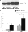
Confirmation of Ref-1 over-expression and the effects of over-expression on TNF-α-inducible IL-8 promoter activity in AGS cells transfected with the −1498/+44 hIL-8/Luc construct. (A) The 37 kDa bands demonstrate increased Ref-1 expression in cells transfected with 0.25 μg pFLAG-Ref-1 cDNA3.1 compared to non-transfected or mock-transfected cells by western blot. (B) The figure depicts the relative increase in normalized IL-8 luciferase activity of treated cells compared to untreated cells with basal levels of Ref-1. The over-expression of Ref-1 significantly (*P < 0.05) increased IL-8 luciferase activity in untreated control cells, and the IL-8 luciferase activity induced by TNF-α in cells with basal levels of Ref-1 was further enhanced in Ref-1 over-expressing cells following TNF-α stimulation (*P < 0.05). Data are expressed as mean relative luciferase activity of the −1498/+44 hIL-8/Luc construct compared to untreated cells with constitutive levels of Ref-1, and results represent the mean ± SEM of three separate experiments.
Fig. 7
Fig. 7
Summary diagram illustrating a model for the mechanism by which Ref-1 regulates TNF-α-induced IL-8 in human gastric epithelial cells. TNF-α acts via its receptor, TNFR-1, on mitochondria to initiate caspase-mediated apoptosis that generates ROS, and/or on IκB kinase to activate NF-κB. ROS themselves can also activate NF-κB, and once activated, free NF-κB translocates into the nucleus. Similarly, either c-Jun/c-Jun homodimers or c-Jun/c-Fos heterodimers are activated by TNF-α or ROS through pathways involving mitogen-activated protein kinases leading to the activation of AP-1. Both ROS and TNF-α increase the expression of Ref-1 in gastric epithelial cells leading to nuclear translocation of Ref-1. The intranuclear presence of Ref-1 reductively modulates AP-1 and NF-κB activation, and in the reduced form, these transcription factors activate transcription by binding to DNA binding sites upstream of several redox-sensitive pro-inflammatory cytokine genes including IL-8.

References

    1. Wiseman H, Halliwell B. Damage to DNA by reactive oxygen and nitrogen species: role in inflammatory disease and progression to cancer. Biochem J. 1996;313:17–29. - PMC - PubMed
    1. Cooper D, Stokes KY, Tailor A, Granger DN. Oxidative stress promotes blood cell–endothelial cell interactions in the microcirculation. Cardiovasc Toxicol. 2002;2(3):165–80. - PubMed
    1. Ding SZ, Minohara Y, Fan XJ, Wang J, Reyes VE, Patel J, et al. Helicobacter pylori infection induces oxidative stress and programmed cell death in human gastric epithelial cells. Infect Immun. 2007;75(8):4030–9. - PMC - PubMed
    1. Crowe SE. Helicobacter infection, chronic inflammation, and the development of malignancy. Curr Opin Gastroenterol. 2005;21(1):32–8. - PubMed
    1. Ernst PB, Peura DA, Crowe SE. The translation of Helicobacter pylori basic research to patient care. Gastroenterology. 2006;130(1):188–206. - PubMed

Publication types

MeSH terms