. 2009 Jul 4;8(7):795-802.
doi: 10.1016/j.dnarep.2009.03.003. Epub 2009 Apr 18.

The role of the retinoblastoma/E2F1 tumor suppressor pathway in the lesion recognition step of nucleotide excision repair

Affiliations

The role of the retinoblastoma/E2F1 tumor suppressor pathway in the lesion recognition step of nucleotide excision repair

Patrick S Lin et al. DNA Repair (Amst). .

Abstract

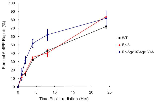
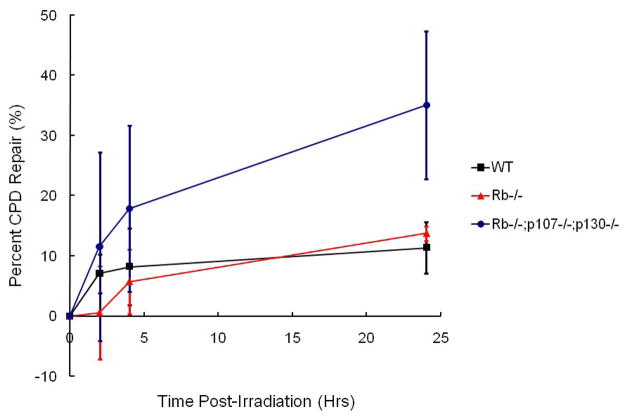
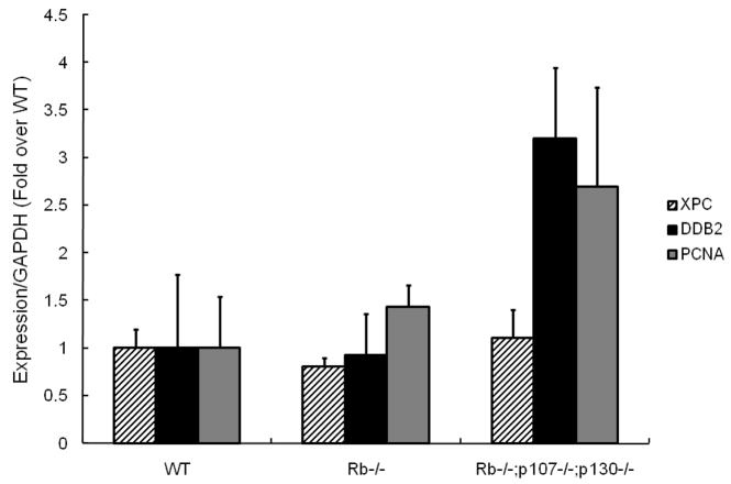
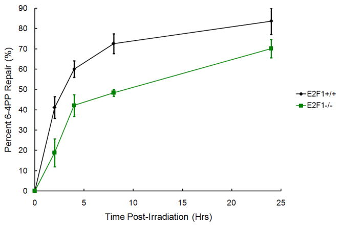
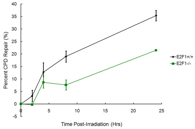
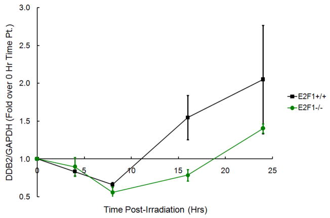
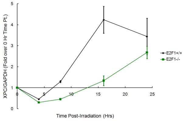
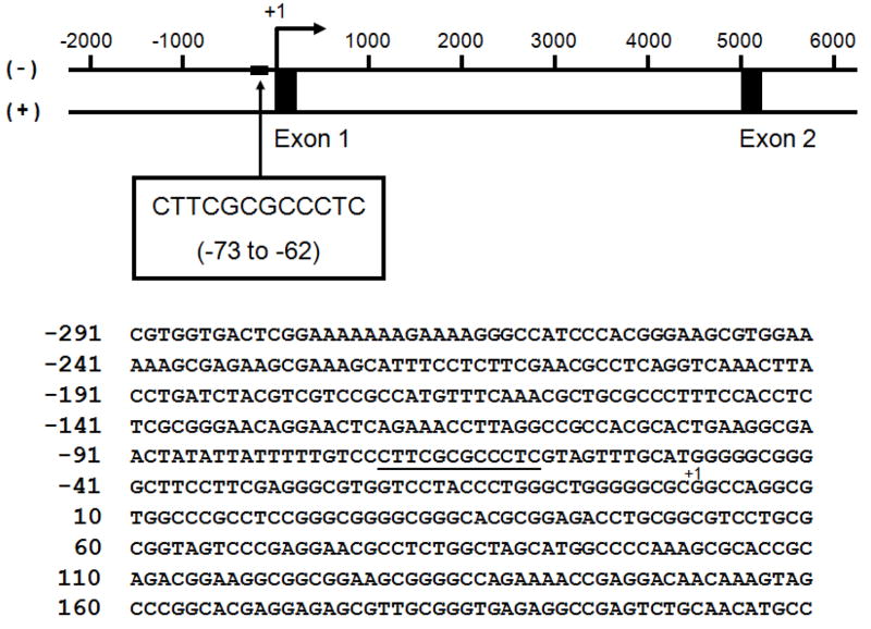
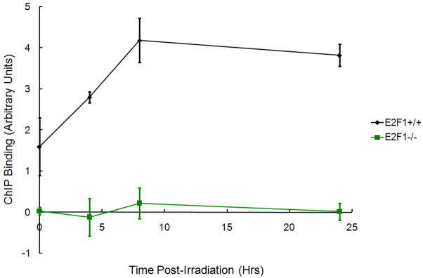
The retinoblastoma Rb/E2F tumor suppressor pathway plays a major role in the regulation of mammalian cell cycle progression. The pRb protein, along with closely related proteins p107 and p130, exerts its anti-proliferative effects by binding to the E2F family of transcription factors known to regulate essential genes throughout the cell cycle. We sought to investigate the role of the Rb/E2F1 pathway in the lesion recognition step of nucleotide excision repair (NER) in mouse embryonic fibroblasts (MEFs). Rb-/-, p107-/-, p130-/- MEFs repaired both cyclobutane pyrimidine dimers (CPDs) and 6-4 photoproducts (6-4PPs) at higher efficiency than did wildtype cells following UV-C irradiation. The expression of damaged DNA binding gene DDB2 involved in the DNA lesion recognition step was elevated in the Rb family-deficient MEFs. To determine if the enhanced DNA repair in the absence of the Rb gene family is due to the derepression of E2F1, we assayed the ability of E2F1-deficient cells to repair damaged DNA and demonstrated that E2F1-/- MEFs are impaired for the removal of both CPDs and 6-4PPs. Furthermore, wildtype cells induced a higher expression of DDB2 and xeroderma pigmentosum gene XPC transcript levels than did E2F1-/- cells following UV-C irradiation. Using an E2F SiteScan algorithm, we uncovered a putative E2F-responsive element in the XPC promoter upstream of the transcription start site. We showed with chromatin immunoprecipitation assays the binding of E2F1 to the XPC promoter in a UV-dependent manner, suggesting that E2F1 is a transcriptional regulator of XPC. Our study identifies a novel E2F1 gene target and further supports the growing body of evidence that the Rb/E2F1 tumor suppressor pathway is involved in the regulation of the DNA lesion recognition step of nucleotide excision repair.


Conflict of interest statement

Conflict of Interest Statement

The authors declare that there is no conflict of interest.

Figures

Figure 1
Figure 1
Global genomic repair assay of wildtype and Rb family-deficient MEFs following 10 J/m2 UV-C irradiation. (a) Repair of 6-4PPs by WT, Rb−/− single mutant, and Rb−/−;p107−/−;p130−/− TKO MEFs. (b) Repair of CPDs by WT, Rb−/− single mutant, and Rb−/−;p107−/−;p130−/− TKO MEFs. Repair curves are represented as percent of DNA repair (y-axis) as a function of time following UV-C irradiation (x-axis). All data points and corresponding standard errors are the result of triplicate experiments (n=3).
Figure 1
Figure 1
Global genomic repair assay of wildtype and Rb family-deficient MEFs following 10 J/m2 UV-C irradiation. (a) Repair of 6-4PPs by WT, Rb−/− single mutant, and Rb−/−;p107−/−;p130−/− TKO MEFs. (b) Repair of CPDs by WT, Rb−/− single mutant, and Rb−/−;p107−/−;p130−/− TKO MEFs. Repair curves are represented as percent of DNA repair (y-axis) as a function of time following UV-C irradiation (x-axis). All data points and corresponding standard errors are the result of triplicate experiments (n=3).
Figure 2
Figure 2
XPC, DDB2, and PCNA transcript levels in wildtype and Rb family-deficient MEFs. Basal, XPC, DDB2, and PCNA transcript levels in WT, Rb−/− single mutant, and Rb−/−;p107−/−;p130−/− TKO MEFs as measured by real time RT-PCR. XPC, DDB2, and PCNA transcript levels are normalized to that of GAPDH and expressed as fold activation over WT level, which is arbitrarily given the value of 1.0. All data points and corresponding standard errors are the result of triplicate experiments (n=3).
Figure 3
Figure 3
Global genomic repair assay of wildtype and E2F1−/− MEFs following 10 J/m2 UV-C irradiation. (a) Repair of 6-4PPs by WT and E2F1−/− MEFs. (b) Repair of CPDs by WT and E2F1−/−MEFs. Repair curves are represented as percent of DNA repair (y-axis) as a function of time following UV-C irradiation (x-axis). All data points and corresponding standard errors are the result of triplicate experiments (n=3).
Figure 3
Figure 3
Global genomic repair assay of wildtype and E2F1−/− MEFs following 10 J/m2 UV-C irradiation. (a) Repair of 6-4PPs by WT and E2F1−/− MEFs. (b) Repair of CPDs by WT and E2F1−/−MEFs. Repair curves are represented as percent of DNA repair (y-axis) as a function of time following UV-C irradiation (x-axis). All data points and corresponding standard errors are the result of triplicate experiments (n=3).
Figure 4
Figure 4
DDB2 and XPC transcript levels in wildtype and E2F1−/− MEFs following 10 J/m2 UV-C irradiation. DNA damage-inducible (a) DDB2 and (b) XPC transcript levels in WT and E2F1−/− MEFs as measured by real time RT-PCR. DDB2 and XPC transcript levels are normalized to that of GAPDH and expressed as fold activation over basal level (0 hour time point), which is arbitrarily given the value of 1.0. All data points and corresponding standard errors are the result of triplicate experiments (n=3).
Figure 4
Figure 4
DDB2 and XPC transcript levels in wildtype and E2F1−/− MEFs following 10 J/m2 UV-C irradiation. DNA damage-inducible (a) DDB2 and (b) XPC transcript levels in WT and E2F1−/− MEFs as measured by real time RT-PCR. DDB2 and XPC transcript levels are normalized to that of GAPDH and expressed as fold activation over basal level (0 hour time point), which is arbitrarily given the value of 1.0. All data points and corresponding standard errors are the result of triplicate experiments (n=3).
Figure 5
Figure 5
A putative E2F1 binding site in the proximal promoter of mouse XPC gene. Schematic diagram showing a putative E2F1 binding site in the mouse XPC gene and the corresponding nucleotide sequence flanking the predicted site (underlined). +1 denotes the transcription start site.
Figure 6
Figure 6
Chromatin immunoprecipitation assay of wildtype and E2F1−/− MEFs following 10 J/m2 UV-C irradiation. The binding of E2F1 to the putative XPC promoter site as measured by real time RT-PCR. All signals are corrected and normalized to levels found in a control site 5000+ base pairs upstream of the putative binding site. All data points and corresponding standard errors are the result of triplicate experiments (n=3).

Similar articles

Cited by

References

    1. Hanawalt PC, Ford JM, Lloyd DR. Functional characterization of global genomic DNA repair and its implications for cancer. Mutat Res. 2003;544:107–114. - PubMed
    1. van Steeg H, Kraemer KH. Xeroderma pigmentosum and the role of UV-induced DNA damage in skin cancer. Mol Med Today. 1999;5:86–94. - PubMed
    1. Wood RD. Nucleotide excision repair in mammalian cells. J Biol Chem. 1997;272:23465–23468. - PubMed
    1. Wood RD, Mitchell M, Sgouros J, Lindahl T. Human DNA repair genes. Science. 2001;291:1284–1289. - PubMed
    1. Bohr VA, Smith CA, Okumoto DS, Hanawalt PC. DNA repair in an active gene: removal of pyrimidine dimers from the DHFR gene of CHO cells is much more efficient than in the genome overall. Cell. 1985;40:359–369. - PubMed

Publication types

MeSH terms

LinkOut - more resources